4WOE

The duplicated taurocyamine kinase from Schistosoma mansoni with bound transition state analog (TSA) components


Experimental Data Snapshot

  • Method: X-RAY DIFFRACTION
  • Resolution: 2.30 Å
  • R-Value Free: 0.256 
  • R-Value Work: 0.185 
  • R-Value Observed: 0.189 

wwPDB Validation   3D Report Full Report


Ligand Structure Quality Assessment 


This is version 1.2 of the entry. See complete history


Literature

The Substrate-free and -bound Crystal Structures of the Duplicated Taurocyamine Kinase from the Human Parasite Schistosoma mansoni.

Merceron, R.Awama, A.M.Montserret, R.Marcillat, O.Gouet, P.

(2015) J Biol Chem 290: 12951-12963

  • DOI: https://doi.org/10.1074/jbc.M114.628909
  • Primary Citation of Related Structures:  
    4WO8, 4WOD, 4WOE

  • PubMed Abstract: 

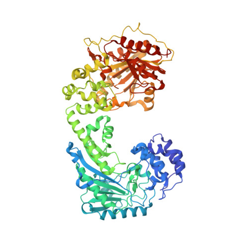
    The taurocyamine kinase from the blood fluke Schistosoma mansoni (SmTK) belongs to the phosphagen kinase (PK) family and catalyzes the reversible Mg(2+)-dependent transfer of a phosphoryl group between ATP and taurocyamine. SmTK is derived from gene duplication, as are all known trematode TKs. Our crystallographic study of SmTK reveals the first atomic structure of both a TK and a PK with a bilobal structure. The two unliganded lobes present a canonical open conformation and interact via their respective C- and N-terminal domains at a helix-mediated interface. This spatial arrangement differs from that observed in true dimeric PKs, in which both N-terminal domains make contact. Our structures of SmTK complexed with taurocyamine or l-arginine compounds explain the mechanism by which an arginine residue of the phosphagen specificity loop is crucial for substrate specificity. An SmTK crystal was soaked with the dead end transition state analog (TSA) components taurocyamine-NO3 (2-)-MgADP. One SmTK monomer was observed with two bound TSAs and an asymmetric conformation, with the first lobe semiclosed and the second closed. However, isothermal titration calorimetry and enzyme kinetics experiments showed that the two lobes function independently. A small angle x-ray scattering model of SmTK-TSA in solution with two closed active sites was generated.


  • Organizational Affiliation

    From the Institut de Biologie et Chimie des Protéines, BMSSI-IBCP, UMR 5086 CNRS Université Lyon 1, 7, Passage du Vercors, 69367 Lyon Cedex 07, France and.


Macromolecules
Find similar proteins by:  (by identity cutoff)  |  3D Structure
Entity ID: 1
MoleculeChains Sequence LengthOrganismDetailsImage
Taurocyamine kinase
A, B
716Schistosoma mansoniMutation(s): 4 
Gene Names: Smp_194770
EC: 2.7.3.4
UniProt
Find proteins for P16641 (Schistosoma mansoni)
Explore P16641 
Go to UniProtKB:  P16641
Entity Groups  
Sequence Clusters30% Identity50% Identity70% Identity90% Identity95% Identity100% Identity
UniProt GroupP16641
Sequence Annotations
Expand
  • Reference Sequence
Small Molecules
Ligands 4 Unique
IDChains Name / Formula / InChI Key2D Diagram3D Interactions
ADP
Query on ADP

Download Ideal Coordinates CCD File 
C [auth A],
D [auth A],
K [auth B],
L [auth B]
ADENOSINE-5'-DIPHOSPHATE
C10 H15 N5 O10 P2
XTWYTFMLZFPYCI-KQYNXXCUSA-N
3S5
Query on 3S5

Download Ideal Coordinates CCD File 
E [auth A],
F [auth A],
M [auth B],
N [auth B]
Taurocyamine
C3 H9 N3 O3 S
JKLRIMRKZBSSED-UHFFFAOYSA-N
NO3
Query on NO3

Download Ideal Coordinates CCD File 
I [auth A],
J [auth A]
NITRATE ION
N O3
NHNBFGGVMKEFGY-UHFFFAOYSA-N
MG
Query on MG

Download Ideal Coordinates CCD File 
G [auth A],
H [auth A],
O [auth B]
MAGNESIUM ION
Mg
JLVVSXFLKOJNIY-UHFFFAOYSA-N
Binding Affinity Annotations 
IDSourceBinding Affinity
ADP Binding MOAD:  4WOE Kd: 3800 (nM) from 1 assay(s)
Experimental Data & Validation

Experimental Data

  • Method: X-RAY DIFFRACTION
  • Resolution: 2.30 Å
  • R-Value Free: 0.256 
  • R-Value Work: 0.185 
  • R-Value Observed: 0.189 
  • Space Group: P 1 21 1
Unit Cell:
Length ( Å )Angle ( ˚ )
a = 107.695α = 90
b = 122.22β = 108.06
c = 63.06γ = 90
Software Package:
Software NamePurpose
PHENIXrefinement
XDSdata reduction
XSCALEdata scaling
PHASERphasing

Structure Validation

View Full Validation Report


Ligand Structure Quality Assessment 


Entry History 

Deposition Data

Revision History  (Full details and data files)

  • Version 1.0: 2015-04-15
    Type: Initial release
  • Version 1.1: 2015-05-27
    Changes: Database references
  • Version 1.2: 2024-01-10
    Changes: Data collection, Database references, Refinement description