4UUY

Structural Identification of the Vps18 beta-propeller reveals a critical role in the HOPS complex stability and function.


Experimental Data Snapshot

  • Method: X-RAY DIFFRACTION
  • Resolution: 2.14 Å
  • R-Value Free: 0.244 
  • R-Value Work: 0.201 
  • R-Value Observed: 0.204 

wwPDB Validation   3D Report Full Report


This is version 1.1 of the entry. See complete history


Literature

Structural Identification of the Vps18 Beta-Propeller Reveals a Critical Role in the Hops Complex Stability and Function.

Behrmann, H.Lurick, A.Kuhlee, A.Balderhaar, H.K.Brocker, C.Kummel, D.Engelbrecht-Vandre, S.Gohlke, U.Raunser, S.Heinemann, U.Ungermann, C.

(2014) J Biol Chem 289: 33503

  • DOI: https://doi.org/10.1074/jbc.M114.602714
  • Primary Citation of Related Structures:  
    4UUY

  • PubMed Abstract: 

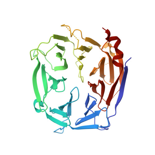
    Membrane fusion at the vacuole, the lysosome equivalent in yeast, requires the HOPS tethering complex, which is recruited by the Rab7 GTPase Ypt7. HOPS provides a template for the assembly of SNAREs and thus likely confers fusion at a distinct position on vacuoles. Five of the six subunits in HOPS have a similar domain prediction with strong similarity to COPII subunits and nuclear porins. Here, we show that Vps18 indeed has a seven-bladed β-propeller as its N-terminal domain by revealing its structure at 2.14 Å. The Vps18 N-terminal domain can interact with the N-terminal part of Vps11 and also binds to lipids. Although deletion of the Vps18 N-terminal domain does not preclude HOPS assembly, as revealed by negative stain electron microscopy, the complex is instable and cannot support membrane fusion in vitro. We thus conclude that the β-propeller of Vps18 is required for HOPS stability and function and that it can serve as a starting point for further structural analyses of the HOPS tethering complex.


  • Organizational Affiliation

    From the Max-Delbrück Center for Molecular Medicine, Macromolecular Structure and Interaction Group, Robert-Rössle-Strasse 10, 13125 Berlin, Germany, Freie Universität Berlin, Chemistry and Biochemistry Institute, Takustrasse 6, 14195 Berlin, Germany.


Macromolecules
Find similar proteins by:  (by identity cutoff)  |  3D Structure
Entity ID: 1
MoleculeChains Sequence LengthOrganismDetailsImage
VACUOLAR MEMBRANE PROTEIN PEP3
A, B
349Saccharomyces cerevisiaeMutation(s): 0 
UniProt
Find proteins for P27801 (Saccharomyces cerevisiae (strain ATCC 204508 / S288c))
Explore P27801 
Go to UniProtKB:  P27801
Entity Groups  
Sequence Clusters30% Identity50% Identity70% Identity90% Identity95% Identity100% Identity
UniProt GroupP27801
Sequence Annotations
Expand
  • Reference Sequence
Small Molecules
Ligands 3 Unique
IDChains Name / Formula / InChI Key2D Diagram3D Interactions
SO4
Query on SO4

Download Ideal Coordinates CCD File 
C [auth A]
D [auth A]
E [auth A]
H [auth B]
I [auth B]
C [auth A],
D [auth A],
E [auth A],
H [auth B],
I [auth B],
J [auth B]
SULFATE ION
O4 S
QAOWNCQODCNURD-UHFFFAOYSA-L
GOL
Query on GOL

Download Ideal Coordinates CCD File 
K [auth B],
L [auth B]
GLYCEROL
C3 H8 O3
PEDCQBHIVMGVHV-UHFFFAOYSA-N
EDO
Query on EDO

Download Ideal Coordinates CCD File 
F [auth A],
G [auth A],
M [auth B]
1,2-ETHANEDIOL
C2 H6 O2
LYCAIKOWRPUZTN-UHFFFAOYSA-N
Experimental Data & Validation

Experimental Data

  • Method: X-RAY DIFFRACTION
  • Resolution: 2.14 Å
  • R-Value Free: 0.244 
  • R-Value Work: 0.201 
  • R-Value Observed: 0.204 
  • Space Group: P 1 21 1
Unit Cell:
Length ( Å )Angle ( ˚ )
a = 56.25α = 90
b = 77.534β = 98.91
c = 84.508γ = 90
Software Package:
Software NamePurpose
REFMACrefinement
XDSdata reduction

Structure Validation

View Full Validation Report


Entry History 

Deposition Data

Revision History  (Full details and data files)

  • Version 1.0: 2014-10-29
    Type: Initial release
  • Version 1.1: 2014-12-17
    Changes: Database references