4TKU | pdb_00004tku

Reactivated Nitrogenase MoFe-protein from A. vinelandii


Experimental Data Snapshot

  • Method: X-RAY DIFFRACTION
  • Resolution: 1.43 Å
  • R-Value Free: 
    0.142 (Depositor), 0.133 (DCC) 
  • R-Value Work: 
    0.132 (Depositor), 0.132 (DCC) 
  • R-Value Observed: 
    0.132 (Depositor) 

wwPDB Validation 3D Report Full Report

Validation slider image for 4TKU

Ligand Structure Quality Assessment 


This is version 1.2 of the entry. See complete history

Literature

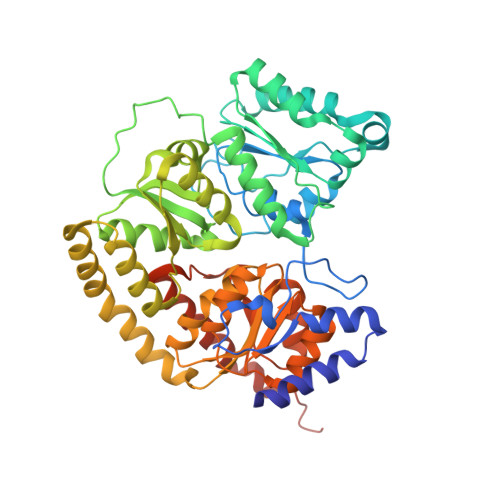
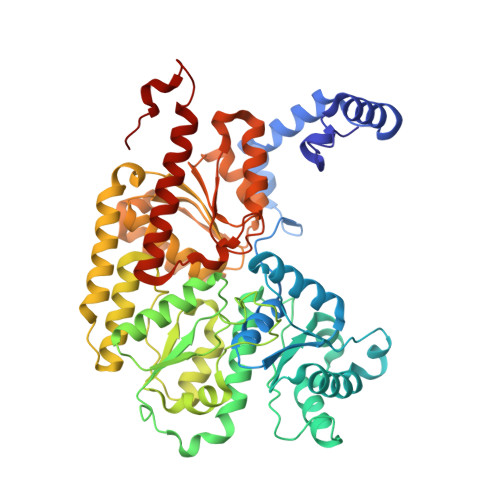
Ligand binding to the FeMo-cofactor: structures of CO-bound and reactivated nitrogenase.

Spatzal, T.Perez, K.A.Einsle, O.Howard, J.B.Rees, D.C.

(2014) Science 345: 1620-1623

  • DOI: https://doi.org/10.1126/science.1256679
  • Primary Citation Related Structures: 
    4TKU, 4TKV

  • PubMed Abstract: 

    The mechanism of nitrogenase remains enigmatic, with a major unresolved issue concerning how inhibitors and substrates bind to the active site. We report a crystal structure of carbon monoxide (CO)-inhibited nitrogenase molybdenum-iron (MoFe)-protein at 1.50 angstrom resolution, which reveals a CO molecule bridging Fe2 and Fe6 of the FeMo-cofactor. The μ2 binding geometry is achieved by replacing a belt-sulfur atom (S2B) and highlights the generation of a reactive iron species uncovered by the displacement of sulfur. The CO inhibition is fully reversible as established by regain of enzyme activity and reappearance of S2B in the 1.43 angstrom resolution structure of the reactivated enzyme. The substantial and reversible reorganization of the FeMo-cofactor accompanying CO binding was unanticipated and provides insights into a catalytically competent state of nitrogenase.


  • Organizational Affiliation
    • Howard Hughes Medical Institute and Division of Chemistry and Chemical Engineering, MailCode 114-96, California Institute of Technology, Pasadena, CA 91125, USA. spatzal@caltech.edu dcrees@caltech.edu.

Macromolecule Content 

  • Total Structure Weight: 233.86 kDa 
  • Atom Count: 17,913 
  • Modeled Residue Count: 1,998 
  • Deposited Residue Count: 2,030 
  • Unique protein chains: 2

Macromolecules

Find similar proteins by:|  3D Structure
Entity ID: 1
MoleculeChains  Sequence LengthOrganismDetailsImage
Nitrogenase molybdenum-iron protein alpha chain
A, C
492Azotobacter vinelandiiMutation(s): 0 
EC: 1.18.6.1
UniProt
Find proteins for P07328 (Azotobacter vinelandii)
Explore P07328 
Go to UniProtKB:  P07328
Entity Groups
Sequence Clusters30% Identity50% Identity70% Identity90% Identity95% Identity100% Identity
UniProt GroupP07328
Sequence Annotations
Expand
Reference Sequence
Find similar proteins by:|  3D Structure
Entity ID: 2
MoleculeChains  Sequence LengthOrganismDetailsImage
Nitrogenase molybdenum-iron protein beta chain
B, D
523Azotobacter vinelandiiMutation(s): 0 
EC: 1.18.6.1
UniProt
Find proteins for P07329 (Azotobacter vinelandii)
Explore P07329 
Go to UniProtKB:  P07329
Entity Groups
Sequence Clusters30% Identity50% Identity70% Identity90% Identity95% Identity100% Identity
UniProt GroupP07329
Sequence Annotations
Expand
Reference Sequence

Small Molecules

Ligands 6 Unique
IDChains Name / Formula / InChI Key2D Diagram3D Interactions
ICS

Query on ICS


Download:Ideal Coordinates CCD File
F [auth A],
O [auth C]
iron-sulfur-molybdenum cluster with interstitial carbon
C Fe7 Mo S9
DDQFAOMIVKLFON-UHFFFAOYSA-N
CLF

Query on CLF


Download:Ideal Coordinates CCD File
I [auth B],
R [auth D]
FE(8)-S(7) CLUSTER
Fe8 S7
JKVMXLBGZBULKV-UHFFFAOYSA-N
HCA

Query on HCA


Download:Ideal Coordinates CCD File
E [auth A],
N [auth C]
3-HYDROXY-3-CARBOXY-ADIPIC ACID
C7 H10 O7
XKJVEVRQMLKSMO-SSDOTTSWSA-N
IMD

Query on IMD


Download:Ideal Coordinates CCD File
G [auth A]
J [auth B]
K [auth B]
L [auth B]
P [auth C]
G [auth A],
J [auth B],
K [auth B],
L [auth B],
P [auth C],
S [auth D],
T [auth D],
U [auth D]
IMIDAZOLE
C3 H5 N2
RAXXELZNTBOGNW-UHFFFAOYSA-O
FE2

Query on FE2


Download:Ideal Coordinates CCD File
M [auth B],
V [auth D]
FE (II) ION
Fe
CWYNVVGOOAEACU-UHFFFAOYSA-N
CL

Query on CL


Download:Ideal Coordinates CCD File
H [auth A],
Q [auth C]
CHLORIDE ION
Cl
VEXZGXHMUGYJMC-UHFFFAOYSA-M

Experimental Data & Validation

Experimental Data

  • Method: X-RAY DIFFRACTION
  • Resolution: 1.43 Å
  • R-Value Free:  0.142 (Depositor), 0.133 (DCC) 
  • R-Value Work:  0.132 (Depositor), 0.132 (DCC) 
  • R-Value Observed: 0.132 (Depositor) 
Space Group: P 1 21 1
Unit Cell:
Length ( Å )Angle ( ˚ )
a = 80.941α = 90
b = 130.785β = 110.58
c = 107.005γ = 90
Software Package:
Software NamePurpose
REFMACrefinement

Structure Validation

View Full Validation Report


Ligand Structure Quality Assessment 


Entry History 

Deposition Data

Revision History  (Full details and data files)

  • Version 1.0: 2014-10-01
    Type: Initial release
  • Version 1.1: 2014-10-15
    Changes: Database references
  • Version 1.2: 2023-12-27
    Changes: Advisory, Data collection, Database references, Derived calculations, Other, Refinement description, Source and taxonomy