4R37

Crystal structure analysis of LpxA, a UDP-N-acetylglucosamine acyltransferase from Bacteroides fragilis 9343 with UDP-GlcNAc


Experimental Data Snapshot

  • Method: X-RAY DIFFRACTION
  • Resolution: 1.90 Å
  • R-Value Free: 0.185 
  • R-Value Work: 0.147 
  • R-Value Observed: 0.149 

wwPDB Validation   3D Report Full Report


Ligand Structure Quality Assessment 


This is version 1.3 of the entry. See complete history


Literature

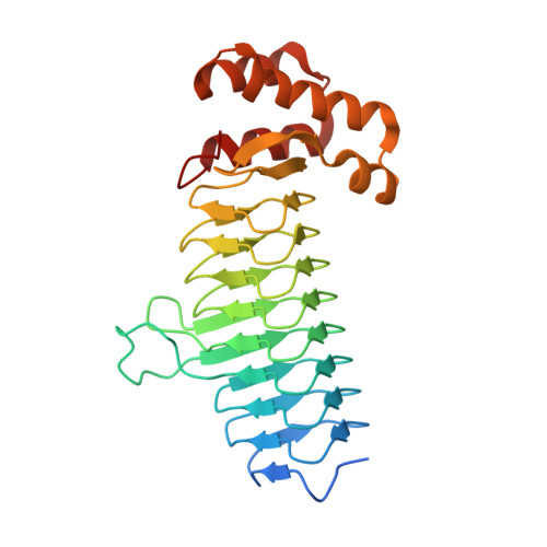
Structures of Bacteroides fragilis uridine 5'-diphosphate-N-acetylglucosamine (UDP-GlcNAc) acyltransferase (BfLpxA).

Ngo, A.Fong, K.T.Cox, D.L.Chen, X.Fisher, A.J.

(2015) Acta Crystallogr D Biol Crystallogr 71: 1068-1076

  • DOI: https://doi.org/10.1107/S1399004715003326
  • Primary Citation of Related Structures:  
    4R36, 4R37

  • PubMed Abstract: 

    Uridine 5'-diphosphate-N-acetylglucosamine (UDP-GlcNAc) acyltransferase (LpxA) catalyzes a reversible reaction for adding an O-acyl group to the GlcNAc in UDP-GlcNAc in the first step of lipid A biosynthesis. Lipid A constitutes a major component of lipopolysaccharides, also referred to as endotoxins, which form the outer monolayer of the outer membrane of Gram-negative bacteria. Ligand-free and UDP-GlcNAc-bound crystal structures of LpxA from Bacteroides fragilis NCTC 9343, the most common pathogenic bacteria found in abdominal abscesses, have been determined and are presented here. The enzyme crystallizes in a cubic space group, with the crystallographic threefold axis generating the biological functional homotrimer and with each monomer forming a nine-rung left-handed β-helical (LβH) fold in the N-terminus followed by an α-helical motif in the C-terminus. The structure is highly similar to LpxA from other organisms. Yet, despite sharing a similar LβH structure with LpxAs from Escherichia coli and others, previously unseen calcium ions are observed on the threefold axis in B. fragilis LpxA to help stabilize the trimeric assembly.


  • Organizational Affiliation

    Department of Chemistry, University of California, One Shields Avenue, Davis, CA 95616, USA.


Macromolecules
Find similar proteins by:  (by identity cutoff)  |  3D Structure
Entity ID: 1
MoleculeChains Sequence LengthOrganismDetailsImage
Putative acyl-[acyl-carrier-protein]--UDP-N-acetylglucosamine O-acyltransferase
A, B
275Bacteroides fragilis NCTC 9343Mutation(s): 0 
Gene Names: lpxABF9343_0789
EC: 2.3.1.129
UniProt
Find proteins for Q5LH16 (Bacteroides fragilis (strain ATCC 25285 / DSM 2151 / CCUG 4856 / JCM 11019 / NCTC 9343 / Onslow))
Explore Q5LH16 
Go to UniProtKB:  Q5LH16
Entity Groups  
Sequence Clusters30% Identity50% Identity70% Identity90% Identity95% Identity100% Identity
UniProt GroupQ5LH16
Sequence Annotations
Expand
  • Reference Sequence
Small Molecules
Ligands 5 Unique
IDChains Name / Formula / InChI Key2D Diagram3D Interactions
UD1
Query on UD1

Download Ideal Coordinates CCD File 
C [auth A]URIDINE-DIPHOSPHATE-N-ACETYLGLUCOSAMINE
C17 H27 N3 O17 P2
LFTYTUAZOPRMMI-CFRASDGPSA-N
PE5
Query on PE5

Download Ideal Coordinates CCD File 
D [auth A]3,6,9,12,15,18,21,24-OCTAOXAHEXACOSAN-1-OL
C18 H38 O9
CUDPPTPIUWYGFI-UHFFFAOYSA-N
PG0
Query on PG0

Download Ideal Coordinates CCD File 
E [auth A]2-(2-METHOXYETHOXY)ETHANOL
C5 H12 O3
SBASXUCJHJRPEV-UHFFFAOYSA-N
ACT
Query on ACT

Download Ideal Coordinates CCD File 
F [auth A],
J [auth B]
ACETATE ION
C2 H3 O2
QTBSBXVTEAMEQO-UHFFFAOYSA-M
CA
Query on CA

Download Ideal Coordinates CCD File 
G [auth A],
H [auth A],
I [auth B],
K [auth B]
CALCIUM ION
Ca
BHPQYMZQTOCNFJ-UHFFFAOYSA-N
Experimental Data & Validation

Experimental Data

  • Method: X-RAY DIFFRACTION
  • Resolution: 1.90 Å
  • R-Value Free: 0.185 
  • R-Value Work: 0.147 
  • R-Value Observed: 0.149 
  • Space Group: P 41 3 2
Unit Cell:
Length ( Å )Angle ( ˚ )
a = 149.403α = 90
b = 149.403β = 90
c = 149.403γ = 90
Software Package:
Software NamePurpose
Blu-Icedata collection
PHASERphasing
REFMACrefinement
XDSdata reduction
Aimlessdata scaling

Structure Validation

View Full Validation Report


Ligand Structure Quality Assessment 


Entry History 

Deposition Data

Revision History  (Full details and data files)

  • Version 1.0: 2015-05-06
    Type: Initial release
  • Version 1.1: 2015-05-20
    Changes: Database references
  • Version 1.2: 2015-06-03
    Changes: Database references
  • Version 1.3: 2023-09-20
    Changes: Data collection, Database references, Derived calculations, Refinement description, Structure summary