4Q49

Room temperature neutron crystal structure of apo human carbonic anhydrase at pH 7.5


Experimental Data Snapshot

  • Method: NEUTRON DIFFRACTION
  • Resolution: 1.80 Å
  • R-Value Free: 0.215 
  • R-Value Work: 0.187 
  • R-Value Observed: 0.190 

wwPDB Validation   3D Report Full Report

Currently 4Q49 does not have a validation slider image.


This is version 1.8 of the entry. See complete history


Literature

Joint neutron crystallographic and NMR solution studies of Tyr residue ionization and hydrogen bonding: Implications for enzyme-mediated proton transfer.

Michalczyk, R.Unkefer, C.J.Bacik, J.P.Schrader, T.E.Ostermann, A.Kovalevsky, A.Y.McKenna, R.Fisher, S.Z.

(2015) Proc Natl Acad Sci U S A 112: 5673-5678

  • DOI: https://doi.org/10.1073/pnas.1502255112
  • Primary Citation of Related Structures:  
    4Q49, 4Y0J

  • PubMed Abstract: 

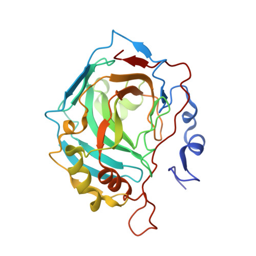
    Human carbonic anhydrase II (HCA II) uses a Zn-bound OH(-)/H2O mechanism to catalyze the reversible hydration of CO2. This catalysis also involves a separate proton transfer step, mediated by an ordered solvent network coordinated by hydrophilic residues. One of these residues, Tyr7, was previously shown to be deprotonated in the neutron crystal structure at pH 10. This observation indicated that Tyr7 has a perturbed pKa compared with free tyrosine. To further probe the pKa of this residue, NMR spectroscopic measurements of [(13)C]Tyr-labeled holo HCA II (with active-site Zn present) were preformed to titrate all Tyr residues between pH 5.4-11.0. In addition, neutron studies of apo HCA II (with Zn removed from the active site) at pH 7.5 and holo HCA II at pH 6 were conducted. This detailed interrogation of tyrosines in HCA II by NMR and neutron crystallography revealed a significantly lowered pKa of Tyr7 and how pH and Tyr proximity to Zn affect hydrogen-bonding interactions.


  • Organizational Affiliation

    Bioscience Division, Los Alamos National Laboratory, Los Alamos, NM 87545;


Macromolecules
Find similar proteins by:  (by identity cutoff)  |  3D Structure
Entity ID: 1
MoleculeChains Sequence LengthOrganismDetailsImage
Carbonic anhydrase 2260Homo sapiensMutation(s): 0 
Gene Names: CA2
EC: 4.2.1.1
UniProt & NIH Common Fund Data Resources
Find proteins for P00918 (Homo sapiens)
Explore P00918 
Go to UniProtKB:  P00918
PHAROS:  P00918
GTEx:  ENSG00000104267 
Entity Groups  
Sequence Clusters30% Identity50% Identity70% Identity90% Identity95% Identity100% Identity
UniProt GroupP00918
Sequence Annotations
Expand
  • Reference Sequence
Small Molecules
Ligands 1 Unique
IDChains Name / Formula / InChI Key2D Diagram3D Interactions
ZN
Query on ZN

Download Ideal Coordinates CCD File 
B [auth A]ZINC ION
Zn
PTFCDOFLOPIGGS-UHFFFAOYSA-N
Experimental Data & Validation

Experimental Data

  • Method: NEUTRON DIFFRACTION
  • Resolution: 1.80 Å
  • R-Value Free: 0.215 
  • R-Value Work: 0.187 
  • R-Value Observed: 0.190 
  • Space Group: P 1 21 1
Unit Cell:
Length ( Å )Angle ( ˚ )
a = 42.78α = 90
b = 41.71β = 104.49
c = 72.82γ = 90
Software Package:
Software NamePurpose
CNSrefinement
nCNSrefinement
PHENIXrefinement
DENZOdata reduction
SCALEPACKdata scaling
CNSphasing

Structure Validation

View Full Validation Report

Currently 4Q49 does not have a validation slider image.


Entry History 

Deposition Data

Revision History  (Full details and data files)

  • Version 1.0: 2015-02-25
    Type: Initial release
  • Version 1.1: 2015-04-22
    Changes: Database references
  • Version 1.2: 2015-05-06
    Changes: Database references
  • Version 1.3: 2015-05-20
    Changes: Database references
  • Version 1.4: 2017-11-22
    Changes: Refinement description
  • Version 1.5: 2018-04-25
    Changes: Data collection
  • Version 1.6: 2018-06-13
    Changes: Data collection
  • Version 1.7: 2021-06-30
    Changes: Data collection, Derived calculations
  • Version 1.8: 2023-09-20
    Changes: Data collection, Database references, Refinement description