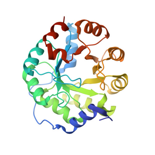
Small molecule probes to quantify the functional fraction of a specific protein in a cell with minimal folding equilibrium shifts.
Liu, Y., Tan, Y.L., Zhang, X., Bhabha, G., Ekiert, D.C., Genereux, J.C., Cho, Y., Kipnis, Y., Bjelic, S., Baker, D., Kelly, J.W.(2014) Proc Natl Acad Sci U S A 111: 4449-4454
- PubMed: 24591605
- DOI: https://doi.org/10.1073/pnas.1323268111
- Primary Citation of Related Structures:
4OU1 - PubMed Abstract:
Although much is known about protein folding in buffers, it remains unclear how the cellular protein homeostasis network functions as a system to partition client proteins between folded and functional, soluble and misfolded, and aggregated conformations. Herein, we develop small molecule folding probes that specifically react with the folded and functional fraction of the protein of interest, enabling fluorescence-based quantification of this fraction in cell lysate at a time point of interest. Importantly, these probes minimally perturb a protein's folding equilibria within cells during and after cell lysis, because sufficient cellular chaperone/chaperonin holdase activity is created by rapid ATP depletion during cell lysis. The folding probe strategy and the faithful quantification of a particular protein's functional fraction are exemplified with retroaldolase, a de novo designed enzyme, and transthyretin, a nonenzyme protein. Our findings challenge the often invoked assumption that the soluble fraction of a client protein is fully folded in the cell. Moreover, our results reveal that the partitioning of destabilized retroaldolase and transthyretin mutants between the aforementioned conformational states is strongly influenced by cytosolic proteostasis network perturbations. Overall, our results suggest that applying a chemical folding probe strategy to other client proteins offers opportunities to reveal how the proteostasis network functions as a system to regulate the folding and function of individual client proteins in vivo.
- Departments of Molecular and Experimental Medicine and Chemistry and The Skaggs Institute for Chemical Biology, The Scripps Research Institute, La Jolla, CA 92037.
Organizational Affiliation:


