4NB4 | pdb_00004nb4

Pantothenamide-bound Pantothenate kinase from Staphylococcus aureus


Experimental Data Snapshot

  • Method: X-RAY DIFFRACTION
  • Resolution: 2.25 Å
  • R-Value Free: 
    0.284 (Depositor), 0.247 (DCC) 
  • R-Value Work: 
    0.230 (Depositor) 
  • R-Value Observed: 
    0.233 (Depositor) 

Starting Model: experimental
View more details

wwPDB Validation 3D Report Full Report

Validation slider image for 4NB4

Ligand Structure Quality Assessment 


This is version 1.2 of the entry. See complete history

Literature

Structural characterization of a new N-substituted pantothenamide bound to pantothenate kinases from Klebsiella pneumoniae and Staphylococcus aureus.

Hughes, S.J.Antoshchenko, T.Kim, K.P.Smil, D.Park, H.W.

(2014) Proteins 82: 1542-1548

  • DOI: https://doi.org/10.1002/prot.24524
  • Primary Citation Related Structures: 
    4NB4, 4NE2

  • PubMed Abstract: 

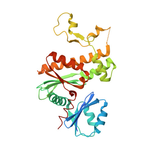
    Pantothenate kinase (PanK) is the rate-limiting enzyme in Coenzyme A biosynthesis, catalyzing the ATP-dependent phosphorylation of pantothenate. We solved the co-crystal structures of PanKs from Staphylococcus aureus (SaPanK) and Klebsiella pneumonia (KpPanK) with N-[2-(1,3-benzodioxol-5-yl)ethyl] pantothenamide (N354-Pan). Two different N354-Pan conformers interact with polar/nonpolar mixed residues in SaPanK and aromatic residues in KpPanK. Additionally, phosphorylated N354-Pan is found at the closed active site of SaPanK but not at the open active site of KpPanK, suggesting an exchange of the phosphorylated product with a new N354-Pan only in KpPanK. Together, pantothenamides conformational flexibility and binding pocket are two key considerations for selective compound design.


  • Organizational Affiliation
    • Department of Pharmacology, University of Toronto, Toronto, Ontario, M5G 1L7.

Macromolecule Content 

  • Total Structure Weight: 257.8 kDa 
  • Atom Count: 17,249 
  • Modeled Residue Count: 2,113 
  • Deposited Residue Count: 2,280 
  • Unique protein chains: 1

Macromolecules

Find similar proteins by:|  3D Structure
Entity ID: 1
MoleculeChains  Sequence LengthOrganismDetailsImage
Type II pantothenate kinase
A, B, C, D, E
A, B, C, D, E, F, G, H
285Staphylococcus aureus subsp. aureus MRSA252Mutation(s): 0 
Gene Names: CoaAcoaW
EC: 2.7.1.33
UniProt
Find proteins for Q6G7I0 (Staphylococcus aureus (strain MSSA476))
Explore Q6G7I0 
Go to UniProtKB:  Q6G7I0
Entity Groups
Sequence Clusters30% Identity50% Identity70% Identity90% Identity95% Identity100% Identity
UniProt GroupQ6G7I0
Sequence Annotations
Expand
Reference Sequence

Small Molecules

Ligands 3 Unique
IDChains Name / Formula / InChI Key2D Diagram3D Interactions
SH3

Query on SH3


Download:Ideal Coordinates CCD File
BA [auth G]
EA [auth H]
J [auth A]
K [auth A]
P [auth C]
BA [auth G],
EA [auth H],
J [auth A],
K [auth A],
P [auth C],
Q [auth C],
X [auth F],
Y [auth F]
N-[2-(1,3-benzodioxol-5-yl)ethyl]-N~3~-[(2R)-2-hydroxy-3,3-dimethyl-4-(phosphonooxy)butanoyl]-beta-alaninamide
C18 H27 N2 O9 P
OQCPPROGRWYIDC-INIZCTEOSA-N
ADP

Query on ADP


Download:Ideal Coordinates CCD File
AA [auth G]
DA [auth H]
I [auth A]
M [auth B]
O [auth C]
AA [auth G],
DA [auth H],
I [auth A],
M [auth B],
O [auth C],
S [auth D],
U [auth E],
W [auth F]
ADENOSINE-5'-DIPHOSPHATE
C10 H15 N5 O10 P2
XTWYTFMLZFPYCI-KQYNXXCUSA-N
MG

Query on MG


Download:Ideal Coordinates CCD File
CA [auth G]
FA [auth H]
L [auth A]
N [auth B]
R [auth C]
CA [auth G],
FA [auth H],
L [auth A],
N [auth B],
R [auth C],
T [auth D],
V [auth E],
Z [auth F]
MAGNESIUM ION
Mg
JLVVSXFLKOJNIY-UHFFFAOYSA-N

Experimental Data & Validation

Experimental Data

  • Method: X-RAY DIFFRACTION
  • Resolution: 2.25 Å
  • R-Value Free:  0.284 (Depositor), 0.247 (DCC) 
  • R-Value Work:  0.230 (Depositor) 
  • R-Value Observed: 0.233 (Depositor) 
Space Group: P 1 21 1
Unit Cell:
Length ( Å )Angle ( ˚ )
a = 103.01α = 90
b = 70.74β = 90.64
c = 143.115γ = 90
Software Package:
Software NamePurpose
SCALAdata scaling
MOLREPphasing
REFMACrefinement
PDB_EXTRACTdata extraction
XDSdata reduction

Structure Validation

View Full Validation Report


Ligand Structure Quality Assessment 


Entry History 

Deposition Data

Revision History  (Full details and data files)

  • Version 1.0: 2014-03-05
    Type: Initial release
  • Version 1.1: 2014-06-25
    Changes: Database references
  • Version 1.2: 2023-09-20
    Changes: Data collection, Database references, Derived calculations, Refinement description