


Crystal structure of a mutant of archaeal ribosomal protein L1 from Methanococcus jannaschii
Sarskikh, A.V., Gabdulkhakov, A.G., Kostareva, O.S., Shkliaeva, A.A., Tishchenko, S.V.(2014) Crystallogr Rep 59: 394-398
Experimental Data Snapshot
Starting Model: experimental
View more details
wwPDB Validation 3D Report Full Report

(2014) Crystallogr Rep 59: 394-398
Entity ID: 1 | |||||
|---|---|---|---|---|---|
| Molecule | Chains | Sequence Length | Organism | Details | Image |
| 50S ribosomal protein L1 | 211 | Methanocaldococcus jannaschii DSM 2661 | Mutation(s): 0 Gene Names: MJ0510, rpl1, rplA | 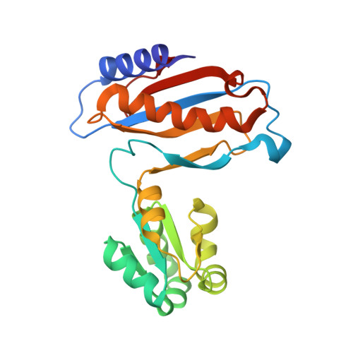 | |
UniProt | |||||
Entity Groups | |||||
| Sequence Clusters | 30% Identity50% Identity70% Identity90% Identity95% Identity100% Identity | ||||
| UniProt Group | P54050 | ||||
Sequence AnnotationsExpand | |||||
Reference Sequence | |||||
| Ligands 3 Unique | |||||
|---|---|---|---|---|---|
| ID | Chains | Name / Formula / InChI Key | 2D Diagram | 3D Interactions | |
| TLA Download:Ideal Coordinates CCD File | B [auth A] | L(+)-TARTARIC ACID C4 H6 O6 FEWJPZIEWOKRBE-JCYAYHJZSA-N |  | ||
| IPA Download:Ideal Coordinates CCD File | C [auth A] | ISOPROPYL ALCOHOL C3 H8 O KFZMGEQAYNKOFK-UHFFFAOYSA-N |  | ||
| CL Download:Ideal Coordinates CCD File | D [auth A], E [auth A], F [auth A] | CHLORIDE ION Cl VEXZGXHMUGYJMC-UHFFFAOYSA-M |  | ||
| Length ( Å ) | Angle ( ˚ ) |
|---|---|
| a = 33.868 | α = 90 |
| b = 105.255 | β = 104.01 |
| c = 38.526 | γ = 90 |
| Software Name | Purpose |
|---|---|
| PROTEUM PLUS | data collection |
| PHASER | phasing |
| PHENIX | refinement |
| PROTEUM PLUS | data reduction |
| PROTEUM PLUS | data scaling |


