


Crystal structure of PHOSPHOTRIESTERASE HOMOLOGY PROTEIN FROM ESCHERICHIA COLI complexed with phosphate in active site
Fedorov, A.A., Fedorov, E.V., Xiang, D.F., Raushel, F.M., Almo, S.C.To be published.
Experimental Data Snapshot
Starting Model: experimental
View more details
Macromolecule Content
Entity ID: 1 | |||||
|---|---|---|---|---|---|
| Molecule | Chains | Sequence Length | Organism | Details | Image |
| Phosphotriesterase homology protein | 292 | Escherichia coli K-12 | Mutation(s): 0 Gene Names: php, yhfV, b3379, JW3342 EC: 3.1 | 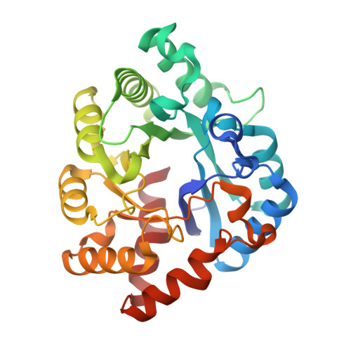 | |
UniProt | |||||
Entity Groups | |||||
| Sequence Clusters | 30% Identity50% Identity70% Identity90% Identity95% Identity100% Identity | ||||
| UniProt Group | P45548 | ||||
Sequence AnnotationsExpand | |||||
Reference Sequence | |||||
| Ligands 3 Unique | |||||
|---|---|---|---|---|---|
| ID | Chains | Name / Formula / InChI Key | 2D Diagram | 3D Interactions | |
| BGC Download:Ideal Coordinates CCD File | GA [auth B] | beta-D-glucopyranose C6 H12 O6 WQZGKKKJIJFFOK-VFUOTHLCSA-N |  | ||
| PO4 Download:Ideal Coordinates CCD File | BA [auth B] BB [auth H] CA [auth B] CB [auth H] DA [auth B] | PHOSPHATE ION O4 P NBIIXXVUZAFLBC-UHFFFAOYSA-K |  | ||
| ZN Download:Ideal Coordinates CCD File | AA [auth B] AB [auth H] HA [auth E] I [auth C] IA [auth E] | ZINC ION Zn PTFCDOFLOPIGGS-UHFFFAOYSA-N |  | ||
| Length ( Å ) | Angle ( ˚ ) |
|---|---|
| a = 83.048 | α = 90 |
| b = 100.308 | β = 104.48 |
| c = 168.037 | γ = 90 |
| Software Name | Purpose |
|---|---|
| CBASS | data collection |
| BALBES | phasing |
| PHENIX | refinement |
| HKL-2000 | data reduction |
| HKL-2000 | data scaling |


