


Crystal structure of a PDZ domain protein (BDI_1242) from Parabacteroides distasonis ATCC 8503 at 2.50 A resolution
Joint Center for Structural Genomics (JCSG)To be published.
Experimental Data Snapshot
wwPDB Validation 3D Report Full Report

Entity ID: 1 | |||||
|---|---|---|---|---|---|
| Molecule | Chains | Sequence Length | Organism | Details | Image |
| PDZ domain protein | 457 | Parabacteroides distasonis ATCC 8503 | Mutation(s): 0 Gene Names: BDI_1242 | 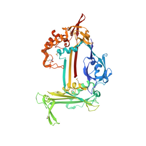 | |
UniProt | |||||
Entity Groups | |||||
| Sequence Clusters | 30% Identity50% Identity70% Identity90% Identity95% Identity100% Identity | ||||
| UniProt Group | A6LBD9 | ||||
Sequence AnnotationsExpand | |||||
Reference Sequence | |||||
| Ligands 3 Unique | |||||
|---|---|---|---|---|---|
| ID | Chains | Name / Formula / InChI Key | 2D Diagram | 3D Interactions | |
| MPD Download:Ideal Coordinates CCD File | C [auth A] | (4S)-2-METHYL-2,4-PENTANEDIOL C6 H14 O2 SVTBMSDMJJWYQN-YFKPBYRVSA-N |  | ||
| EOH Download:Ideal Coordinates CCD File | B [auth A] | ETHANOL C2 H6 O LFQSCWFLJHTTHZ-UHFFFAOYSA-N |  | ||
| MG Download:Ideal Coordinates CCD File | D [auth A] | MAGNESIUM ION Mg JLVVSXFLKOJNIY-UHFFFAOYSA-N |  | ||
| Modified Residues 1 Unique | |||||
|---|---|---|---|---|---|
| ID | Chains | Type | Formula | 2D Diagram | Parent |
| MSE Query on MSE | A | L-PEPTIDE LINKING | C5 H11 N O2 Se |  | MET |
| Length ( Å ) | Angle ( ˚ ) |
|---|---|
| a = 104.093 | α = 90 |
| b = 104.093 | β = 90 |
| c = 96.277 | γ = 120 |
| Software Name | Purpose |
|---|---|
| MolProbity | model building |
| PDB_EXTRACT | data extraction |
| SHELX | phasing |
| SHARP | phasing |
| XSCALE | data scaling |
| BUSTER-TNT | refinement |
| XDS | data reduction |
| SHELXD | phasing |
| BUSTER | refinement |


