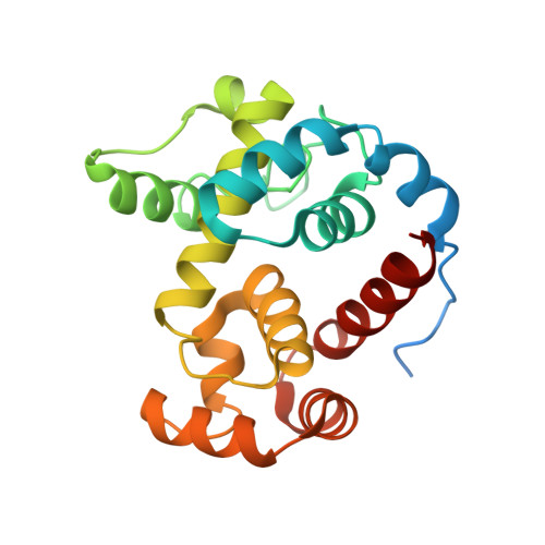
4HJY

2.4 A Crystal structure of E. coli MltE-E64Q with bound chitopentaose


Experimental Data Snapshot

  • Method: X-RAY DIFFRACTION
  • Resolution: 2.40 Å
  • R-Value Free: 0.261 
  • R-Value Work: 0.198 
  • R-Value Observed: 0.204 

wwPDB Validation   3D Report Full Report


This is version 2.1 of the entry. See complete history


Literature

On the Mechanism of Peptidoglycan Binding and Cleavage by the endo-Specific Lytic Transglycosylase MltE from Escherichia coli.

Fibriansah, G.Gliubich, F.I.Thunnissen, A.M.

(2012) Biochemistry 51: 9164-9177

  • DOI: https://doi.org/10.1021/bi300900t
  • Primary Citation of Related Structures:  
    3T36, 4HJV, 4HJY, 4HJZ

  • PubMed Abstract: 

    The lytic transglycosylase MltE from Escherichia coli is a periplasmic, outer membrane-attached enzyme that cleaves the β-1,4-glycosidic bonds between N-acetylmuramic acid and N-acetylglucosamine residues in the cell wall peptidoglycan, producing 1,6-anhydromuropeptides. Here we report three crystal structures of MltE: in a substrate-free state, in a binary complex with chitopentaose, and in a ternary complex with the glycopeptide inhibitor bulgecin A and the murodipeptide N-acetylglucosaminyl-N-acetylmuramyl-l-Ala-d-Glu. The substrate-bound structures allowed a detailed analysis of the saccharide-binding interactions in six subsites of the peptidoglycan-binding groove (subsites -4 to +2) and, combined with site-directed mutagenesis analysis, confirmed the role of Glu64 as catalytic acid/base. The structures permitted the precise modeling of a short glycan strand of eight saccharide residues, providing evidence for two additional subsites (+3 and +4) and revealing the productive conformational state of the substrate at subsites -1 and +1, where the glycosidic bond is cleaved. Full accessibility of the peptidoglycan-binding groove and preferential binding of an N-acetylmuramic acid residue in a (4)C(1) chair conformation at subsite +2 explain why MltE shows only endo- and no exo-specific activity toward glycan strands. The results further indicate that catalysis of glycosidic bond cleavage by MltE proceeds via distortion toward a sofa-like conformation of the N-acetylmuramic acid sugar ring at subsite -1 and by anchimeric assistance of the sugar's N-acetyl group, as shown previously for the lytic transglycosylases Slt70 and MltB.


  • Organizational Affiliation

    Laboratory of Biophysical Chemistry, Groningen Biomolecular Sciences and Biotechnology Institute, University of Groningen, Nijenborgh 7, 9747 AG Groningen, The Netherlands.


Macromolecules
Find similar proteins by:  (by identity cutoff)  |  3D Structure
Entity ID: 1
MoleculeChains Sequence LengthOrganismDetailsImage
Endo-type membrane-bound lytic murein transglycosylase A
A, B
206Escherichia coli K-12Mutation(s): 1 
Gene Names: emtAmltEsltZycgPb1193JW5821
EC: 4.2.2
UniProt
Find proteins for P0C960 (Escherichia coli (strain K12))
Explore P0C960 
Go to UniProtKB:  P0C960
Entity Groups  
Sequence Clusters30% Identity50% Identity70% Identity90% Identity95% Identity100% Identity
UniProt GroupP0C960
Sequence Annotations
Expand
  • Reference Sequence
Oligosaccharides

Help

Entity ID: 2
MoleculeChains Length2D Diagram Glycosylation3D Interactions
2-acetamido-2-deoxy-beta-D-glucopyranose-(1-4)-2-acetamido-2-deoxy-beta-D-glucopyranose-(1-4)-2-acetamido-2-deoxy-beta-D-glucopyranose-(1-4)-2-acetamido-2-deoxy-beta-D-glucopyranose-(1-4)-2-acetamido-2-deoxy-beta-D-glucopyranose
C
5N/A
Glycosylation Resources
GlyTouCan:  G07670VS
GlyCosmos:  G07670VS
GlyGen:  G07670VS
Entity ID: 3
MoleculeChains Length2D Diagram Glycosylation3D Interactions
2-acetamido-2-deoxy-beta-D-glucopyranose-(1-4)-2-acetamido-2-deoxy-beta-D-glucopyranose-(1-4)-2-acetamido-2-deoxy-beta-D-glucopyranose
D
3N/A
Glycosylation Resources
GlyTouCan:  G47362BJ
GlyCosmos:  G47362BJ
GlyGen:  G47362BJ
Biologically Interesting Molecules (External Reference) 1 Unique
Experimental Data & Validation

Experimental Data

  • Method: X-RAY DIFFRACTION
  • Resolution: 2.40 Å
  • R-Value Free: 0.261 
  • R-Value Work: 0.198 
  • R-Value Observed: 0.204 
  • Space Group: P 1 21 1
Unit Cell:
Length ( Å )Angle ( ˚ )
a = 71.54α = 90
b = 33.62β = 106.05
c = 75.22γ = 90
Software Package:
Software NamePurpose
MOSFLMdata reduction
SCALAdata scaling
MOLREPphasing
PHENIXrefinement
PDB_EXTRACTdata extraction
MxCuBEdata collection

Structure Validation

View Full Validation Report


Entry History 

Deposition Data

Revision History  (Full details and data files)

  • Version 1.0: 2012-10-24
    Type: Initial release
  • Version 1.1: 2012-10-31
    Changes: Database references
  • Version 1.2: 2012-12-26
    Changes: Database references
  • Version 2.0: 2020-07-29
    Type: Remediation
    Reason: Carbohydrate remediation
    Changes: Atomic model, Data collection, Database references, Derived calculations, Structure summary
  • Version 2.1: 2023-09-20
    Changes: Data collection, Database references, Refinement description, Structure summary