Potent inhibition of mandelate racemase by a fluorinated substrate-product analogue with a novel binding mode.
Nagar, M., Lietzan, A.D., St Maurice, M., Bearne, S.L.(2014) Biochemistry 53: 1169-1178
- PubMed: 24472022 Search on PubMed
- DOI: https://doi.org/10.1021/bi401703h
- Primary Citation Related Structures:
4FP1, 4HNC, 4M6U - PubMed Abstract:
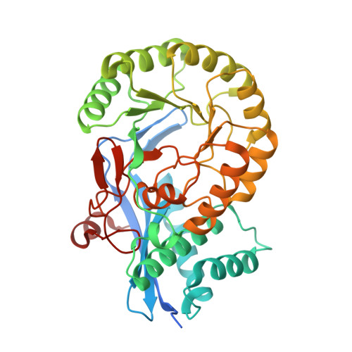
Mandelate racemase (MR) from Pseudomonas putida catalyzes the Mg(2+)-dependent 1,1-proton transfer that interconverts the enantiomers of mandelate. Because trifluorolactate is also a substrate of MR, we anticipated that replacing the phenyl rings of the competitive, substrate-product analogue inhibitor benzilate (Ki = 0.7 mM) with trifluoromethyl groups might furnish an inhibitor. Surprisingly, the substrate-product analogue 3,3,3-trifluoro-2-hydroxy-2-(trifluoromethyl)propanoate (TFHTP) was a potent competitive inhibitor [Ki = 27 ± 4 μM; cf. Km = 1.2 mM for both (R)-mandelate and (R)-trifluorolactate]. To understand the origins of this high binding affinity, we determined the X-ray crystal structure of the MR-TFHTP complex to 1.68 Å resolution. Rather than chelating the active site Mg(2+) with its glycolate moiety, like other ground state analogues, TFHTP exhibited a novel binding mode with the two trifluoromethyl groups closely packed against the 20s loop and the carboxylate bridging the two active site Brønsted acid-base catalysts Lys 166 and His 297. Recognizing that positioning a carboxylate between the Brønsted acid-base catalysts could yield an inhibitor, we showed that tartronate was a competitive inhibitor of MR (Ki = 1.8 ± 0.1 mM). The X-ray crystal structure of the MR-tartronate complex (1.80 Å resolution) revealed that the glycolate moiety of tartronate chelated the Mg(2+) and that the carboxylate bridged Lys 166 and His 297. Models of tartronate in monomers A and B of the crystal structure mimicked the binding orientations of (S)-mandelate and that anticipated for (R)-mandelate, respectively. For the latter monomer, the 20s loop appeared to be disordered, as it also did in the X-ray structure of the MR triple mutant (C92S/C264S/K166C) complexed with benzilate, which was determined to 1.89 Å resolution. These observations indicate that the 20s loop likely undergoes a significant conformational change upon binding (R)-mandelate. In general, our observations suggest that inhibitors of other enolase superfamily enzymes may be designed to capitalize on the recognition of the active site Brønsted acid-base catalysts as binding determinants.
- Department of Biochemistry and Molecular Biology, Dalhousie University , Halifax, Nova Scotia B3H 4R2, Canada.
Organizational Affiliation: