4FLN

Crystal structure of plant protease Deg2


Experimental Data Snapshot

  • Method: X-RAY DIFFRACTION
  • Resolution: 2.80 Å
  • R-Value Free: 0.274 
  • R-Value Work: 0.203 
  • R-Value Observed: 0.207 

wwPDB Validation   3D Report Full Report


This is version 1.2 of the entry. See complete history


Literature

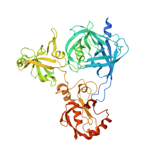
Crystal structure of Arabidopsis deg2 protein reveals an internal PDZ ligand locking the hexameric resting state.

Sun, R.Fan, H.Gao, F.Lin, Y.Zhang, L.Gong, W.Liu, L.

(2012) J Biol Chem 287: 37564-37569

  • DOI: https://doi.org/10.1074/jbc.M112.394585
  • Primary Citation of Related Structures:  
    4FLN

  • PubMed Abstract: 

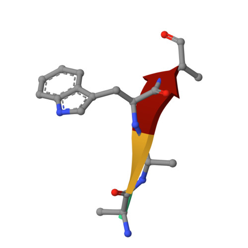
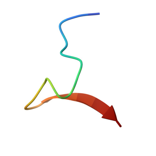
    Eukaryotic organelles have developed elaborate protein quality control systems to ensure their normal activity, among which Deg/HtrA proteases play an essential role. Plant Deg2 protease is a homologue of prokaryotic DegQ/DegP proteases and is located in the chloroplast stroma, where its proteolytic activity is required to maintain the efficiency of photosynthetic machinery during stress. Here, we demonstrate that Deg2 exhibits dual protease-chaperone activities, and we present the hexameric structure of Deg2 complexed with co-purified peptides. The structure shows that Deg2 contains a unique second PDZ domain (PDZ2) following a conventional PDZ domain (PDZ1), with PDZ2 orchestrating the cage assembly of Deg2. We discovered a conserved internal ligand for PDZ2 that mediates hexamer formation and thus locks the protease in the resting state. These findings provide insight into the diverse modes of PDZ domain-mediated regulation of Deg proteases.


  • Organizational Affiliation

    Photosynthesis Research Center, Key Laboratory of Photobiology, Institute of Botany, Chinese Academy of Sciences, Beijing 100093, China.


Macromolecules
Find similar proteins by:  (by identity cutoff)  |  3D Structure
Entity ID: 1
MoleculeChains Sequence LengthOrganismDetailsImage
Protease Do-like 2, chloroplastic
A, B, C
539Arabidopsis thalianaMutation(s): 0 
Gene Names: DEGP2At2g47940F17A22.33T9J23.7
EC: 3.4.21
UniProt
Find proteins for O82261 (Arabidopsis thaliana)
Explore O82261 
Go to UniProtKB:  O82261
Entity Groups  
Sequence Clusters30% Identity50% Identity70% Identity90% Identity95% Identity100% Identity
UniProt GroupO82261
Sequence Annotations
Expand
  • Reference Sequence

Find similar proteins by:  Sequence   |   3D Structure  

Entity ID: 2
MoleculeChains Sequence LengthOrganismDetailsImage
Unknown peptide
D, F
4unidentifiedMutation(s): 0 
Sequence Annotations
Expand
  • Reference Sequence

Find similar proteins by:  Sequence   |   3D Structure  

Entity ID: 3
MoleculeChains Sequence LengthOrganismDetailsImage
Unknown peptide20unidentifiedMutation(s): 0 
Entity Groups  
Sequence Clusters30% Identity50% Identity70% Identity90% Identity95% Identity100% Identity
Sequence Annotations
Expand
  • Reference Sequence
Experimental Data & Validation

Experimental Data

  • Method: X-RAY DIFFRACTION
  • Resolution: 2.80 Å
  • R-Value Free: 0.274 
  • R-Value Work: 0.203 
  • R-Value Observed: 0.207 
  • Space Group: C 2 2 21
Unit Cell:
Length ( Å )Angle ( ˚ )
a = 125.398α = 90
b = 188.165β = 90
c = 166.131γ = 90
Software Package:
Software NamePurpose
HKL-2000data collection
SHELXSphasing
PHENIXrefinement
HKL-2000data reduction
HKL-2000data scaling

Structure Validation

View Full Validation Report


Entry History 

Deposition Data

Revision History  (Full details and data files)

  • Version 1.0: 2012-09-19
    Type: Initial release
  • Version 1.1: 2012-12-19
    Changes: Database references
  • Version 1.2: 2024-02-28
    Changes: Data collection, Database references