4BKF

crystal structure of the human EphA4 ectodomain in complex with human ephrinB3


Experimental Data Snapshot

  • Method: X-RAY DIFFRACTION
  • Resolution: 4.65 Å
  • R-Value Free: 0.351 
  • R-Value Work: 0.327 
  • R-Value Observed: 0.329 

wwPDB Validation   3D Report Full Report


This is version 1.5 of the entry. See complete history


Literature

Structurally Encoded Intraclass Differences in Epha Clusters Drive Distinct Cell Responses

Seiradake, E.Schaupp, A.Del Toro Ruiz, D.Kaufmann, R.Mitakidis, N.Harlos, K.Aricescu, A.R.Klein, R.Jones, E.Y.

(2013) Nat Struct Mol Biol 20: 958

  • DOI: https://doi.org/10.1038/nsmb.2617
  • Primary Citation of Related Structures:  
    4BK4, 4BK5, 4BKA, 4BKF

  • PubMed Abstract: 

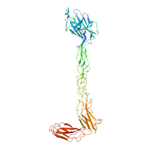
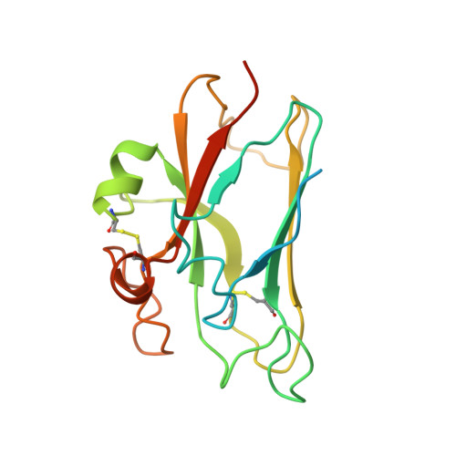
    Functional outcomes of ephrin binding to Eph receptors (Ephs) range from cell repulsion to adhesion. Here we used cell collapse and stripe assays, showing contrasting effects of human ephrinA5 binding to EphA2 and EphA4. Despite equivalent ligand binding affinities, EphA4 triggered greater cell collapse, whereas EphA2-expressing cells adhered better to ephrinA5-coated surfaces. Chimeric receptors showed that the ectodomain is a major determinant of cell response. We report crystal structures of EphA4 ectodomain alone and in complexes with ephrinB3 and ephrinA5. These revealed closed clusters with a dimeric or circular arrangement in the crystal lattice, contrasting with extended arrays previously observed for EphA2 ectodomain. Localization microscopy showed that ligand-stimulated EphA4 induces smaller clusters than does EphA2. Mutant Ephs link these characteristics to interactions observed in the crystal lattices, suggesting a mechanism by which distinctive ectodomain surfaces determine clustering, and thereby signaling, properties.


  • Organizational Affiliation

    Division of Structural Biology, Wellcome Trust Centre for Human Genetics, University of Oxford, Oxford, UK.


Macromolecules
Find similar proteins by:  (by identity cutoff)  |  3D Structure
Entity ID: 1
MoleculeChains Sequence LengthOrganismDetailsImage
EPHRIN TYPE-A RECEPTOR 4
A, B
568Homo sapiensMutation(s): 0 
EC: 2.7.10.1
UniProt & NIH Common Fund Data Resources
Find proteins for P54764 (Homo sapiens)
Explore P54764 
Go to UniProtKB:  P54764
PHAROS:  P54764
GTEx:  ENSG00000116106 
Entity Groups  
Sequence Clusters30% Identity50% Identity70% Identity90% Identity95% Identity100% Identity
UniProt GroupP54764
Sequence Annotations
Expand
  • Reference Sequence
Find similar proteins by:  (by identity cutoff)  |  3D Structure
Entity ID: 2
MoleculeChains Sequence LengthOrganismDetailsImage
EPHRIN-B3
C, D
183Homo sapiensMutation(s): 0 
UniProt & NIH Common Fund Data Resources
Find proteins for Q15768 (Homo sapiens)
Explore Q15768 
Go to UniProtKB:  Q15768
PHAROS:  Q15768
GTEx:  ENSG00000108947 
Entity Groups  
Sequence Clusters30% Identity50% Identity70% Identity90% Identity95% Identity100% Identity
UniProt GroupQ15768
Sequence Annotations
Expand
  • Reference Sequence
Experimental Data & Validation

Experimental Data

  • Method: X-RAY DIFFRACTION
  • Resolution: 4.65 Å
  • R-Value Free: 0.351 
  • R-Value Work: 0.327 
  • R-Value Observed: 0.329 
  • Space Group: P 43 3 2
Unit Cell:
Length ( Å )Angle ( ˚ )
a = 300.53α = 90
b = 300.53β = 90
c = 300.53γ = 90
Software Package:
Software NamePurpose
BUSTERrefinement
XDSdata reduction
XSCALEdata scaling
PHASERphasing

Structure Validation

View Full Validation Report


Entry History 

Deposition Data

Revision History  (Full details and data files)

  • Version 1.0: 2013-07-03
    Type: Initial release
  • Version 1.1: 2013-07-10
    Changes: Database references
  • Version 1.2: 2013-08-21
    Changes: Database references
  • Version 1.3: 2015-11-11
    Changes: Data collection
  • Version 1.4: 2019-04-03
    Changes: Data collection, Other, Source and taxonomy
  • Version 1.5: 2023-12-20
    Changes: Data collection, Database references, Other, Refinement description