4AUO | pdb_00004auo

Crystal structure of MMP-1(E200A) in complex with a triple-helical collagen peptide


Experimental Data Snapshot

  • Method: X-RAY DIFFRACTION
  • Resolution: 3.00 Å
  • R-Value Free: 
    0.273 (Depositor), 0.274 (DCC) 
  • R-Value Work: 
    0.211 (Depositor), 0.212 (DCC) 
  • R-Value Observed: 
    0.211 (Depositor) 

Starting Model: experimental
View more details

wwPDB Validation 3D Report Full Report

Validation slider image for 4AUO

This is version 1.2 of the entry. See complete history

Literature

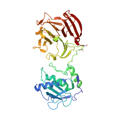
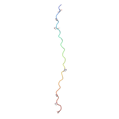
Structural Insights Into Triple-Helical Collagen Cleavage by Matrix Metalloproteinase 1

Manka, S.W.Carafoli, F.Visse, R.Bihan, D.Raynal, N.Farndale, R.W.Murphy, G.Enghild, J.J.Hohenester, E.Nagase, H.

(2012) Proc Natl Acad Sci U S A 109: 12461

  • DOI: https://doi.org/10.1073/pnas.1204991109
  • Primary Citation Related Structures: 
    4AUO

  • PubMed Abstract: 

    Collagenases of the matrix metalloproteinase (MMP) family play major roles in morphogenesis, tissue repair, and human diseases, but how they recognize and cleave the collagen triple helix is not fully understood. Here, we report temperature-dependent binding of a catalytically inactive MMP-1 mutant (E200A) to collagen through the cooperative action of its catalytic and hemopexin domains. Contact between the two molecules was mapped by screening the Collagen Toolkit peptide library and by hydrogen/deuterium exchange. The crystal structure of MMP-1(E200A) bound to a triple-helical collagen peptide revealed extensive interactions of the 115-Å-long triple helix with both MMP-1 domains. An exosite in the hemopexin domain, which binds the leucine 10 residues C-terminal to the scissile bond, is critical for collagenolysis and represents a unique target for inhibitor development. The scissile bond is not correctly positioned for hydrolysis in the crystallized complex. A productive binding mode is readily modeled, without altering the MMP-1 structure or the exosite interactions, by axial rotation of the collagen homotrimer. Interdomain flexing of the enzyme and a localized excursion of the collagen chain closest to the active site, facilitated by thermal loosening of the substrate, may lead to the first transition state of collagenolysis.


  • Organizational Affiliation
    • Nuffield Department of Orthopaedics, Rheumatology and Musculoskeletal Sciences, Kennedy Institute of Rheumatology, University of Oxford, London W6 8LH, United Kingdom.

Macromolecule Content 

  • Total Structure Weight: 107.74 kDa 
  • Atom Count: 7,387 
  • Modeled Residue Count: 947 
  • Deposited Residue Count: 974 
  • Unique protein chains: 2

Macromolecules

Find similar proteins by:|  3D Structure
Entity ID: 1
MoleculeChains  Sequence LengthOrganismDetailsImage
INTERSTITIAL COLLAGENASE
A, B
367Homo sapiensMutation(s): 1 
EC: 3.4.24.7
UniProt & NIH Common Fund Data Resources
Find proteins for P03956 (Homo sapiens)
Explore P03956 
Go to UniProtKB:  P03956
PHAROS:  P03956
GTEx:  ENSG00000196611 
Entity Groups
Sequence Clusters30% Identity50% Identity70% Identity90% Identity95% Identity100% Identity
UniProt GroupP03956
Sequence Annotations
Expand
Reference Sequence
Find similar proteins by:|  3D Structure
Entity ID: 2
MoleculeChains  Sequence LengthOrganismDetailsImage
TRIPLE-HELICAL COLLAGEN PEPTIDE
C, D, E, F, G
C, D, E, F, G, H
40synthetic constructMutation(s): 0 

Small Molecules

Ligands 2 Unique
IDChains Name / Formula / InChI Key2D Diagram3D Interactions
ZN

Query on ZN


Download:Ideal Coordinates CCD File
M [auth A],
N [auth A],
S [auth B],
T [auth B]
ZINC ION
Zn
PTFCDOFLOPIGGS-UHFFFAOYSA-N
CA

Query on CA


Download:Ideal Coordinates CCD File
I [auth A]
J [auth A]
K [auth A]
L [auth A]
O [auth B]
I [auth A],
J [auth A],
K [auth A],
L [auth A],
O [auth B],
P [auth B],
Q [auth B],
R [auth B]
CALCIUM ION
Ca
BHPQYMZQTOCNFJ-UHFFFAOYSA-N
Modified Residues  1 Unique
IDChains TypeFormula2D DiagramParent
HYP
Query on HYP
C, D, E, F, G
C, D, E, F, G, H
L-PEPTIDE LINKINGC5 H9 N O3PRO

Experimental Data & Validation

Experimental Data

  • Method: X-RAY DIFFRACTION
  • Resolution: 3.00 Å
  • R-Value Free:  0.273 (Depositor), 0.274 (DCC) 
  • R-Value Work:  0.211 (Depositor), 0.212 (DCC) 
  • R-Value Observed: 0.211 (Depositor) 
Space Group: P 1 2 1
Unit Cell:
Length ( Å )Angle ( ˚ )
a = 76.671α = 90
b = 102.241β = 103.75
c = 80.734γ = 90
Software Package:
Software NamePurpose
CNSrefinement
MOSFLMdata reduction
SCALAdata scaling
PHASERphasing

Structure Validation

View Full Validation Report


Entry History 

Deposition Data

Revision History  (Full details and data files)

  • Version 1.0: 2012-07-11
    Type: Initial release
  • Version 1.1: 2012-08-15
    Changes: Database references
  • Version 1.2: 2023-12-20
    Changes: Data collection, Database references, Derived calculations, Other, Refinement description