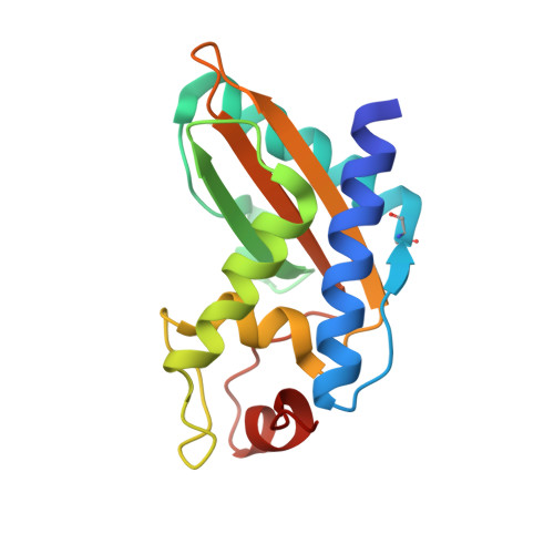
4AIW | pdb_00004aiw

GAPR-1 with bound inositol hexakisphosphate


Experimental Data Snapshot

  • Method: X-RAY DIFFRACTION
  • Resolution: 1.50 Å
  • R-Value Free: 
    0.187 (Depositor), 0.200 (DCC) 
  • R-Value Work: 
    0.152 (Depositor), 0.170 (DCC) 
  • R-Value Observed: 
    0.154 (Depositor) 

Starting Model: experimental
View more details

wwPDB Validation   3D Report Full Report


Ligand Structure Quality Assessment 

Created with Raphaël 2.3.0Worse 01 BetterLigand structure goodness of fit to experimental dataBest fitted IHPClick on this verticalbar to view details

This is version 2.1 of the entry. See complete history


Literature

Interaction of Gapr-1 with Lipid Bilayers is Regulated by Alternative Homodimerization.

Van Galen, J.Olrichs, N.K.Schouten, A.Serrano, R.L.Nolte-'T Hoen, E.N.M.Eerland, R.Kaloyanova, D.Gros, P.Helms, J.B.

(2012) Biochim Biophys Acta 1818: 2175

  • DOI: https://doi.org/10.1016/j.bbamem.2012.04.016
  • Primary Citation of Related Structures:  
    4AIW

  • PubMed Abstract: 

    Golgi-Associated Plant Pathogenesis-Related protein 1 (GAPR-1) is a mammalian protein that belongs to the superfamily of plant pathogenesis related proteins group 1 (PR-1). GAPR-1 is a peripheral membrane-binding protein that strongly associates with lipid-enriched microdomains at the cytosolic leaflet of Golgi membranes. Little is known about the mechanism of GAPR-1 interaction with membranes. We previously suggested that dimerization plays a role in the function of GAPR-1 and here we report that phytic acid (inositol hexakisphosphate) induces dimerization of GAPR-1 in solution. Elucidation of the crystal structure of GAPR-1 in the presence of phytic acid revealed that the GAPR-1 dimer differs from the previously published GAPR-1 dimer structure. In this structure, one of the monomeric subunits of the crystallographic dimer is rotated by 28.5°. To study the GAPR-1 dimerization properties, we investigated the interaction with liposomes in a light scattering assay and by flow cytometry. In the presence of negatively charged lipids, GAPR-1 caused a rapid and stable tethering of liposomes. [D81K]GAPR-1, a mutant predicted to stabilize the IP6-induced dimer conformation, also caused tethering of liposomes. [A68K]GAPR-1 however, a mutant predicted to stabilize the non-rotated dimer conformation, is capable of binding to liposomes but did not cause liposome tethering. Our combined data suggest that the charge properties of the lipid bilayer can regulate GAPR-1 dynamics as a potential mechanism to modulate GAPR-1 function.


  • Organizational Affiliation

    Department of Biochemistry and Cell Biology, Utrecht University, Utrecht, The Netherlands.


Macromolecules
Find similar proteins by:  (by identity cutoff)  |  3D Structure
Entity ID: 1
MoleculeChains Sequence LengthOrganismDetailsImage
GOLGI-ASSOCIATED PLANT PATHOGENESIS-RELATED PROTEIN 1154Homo sapiensMutation(s): 0 
UniProt & NIH Common Fund Data Resources
Find proteins for Q9H4G4 (Homo sapiens)
Explore Q9H4G4 
Go to UniProtKB:  Q9H4G4
PHAROS:  Q9H4G4
GTEx:  ENSG00000122694 
Entity Groups  
Sequence Clusters30% Identity50% Identity70% Identity90% Identity95% Identity100% Identity
UniProt GroupQ9H4G4
Sequence Annotations
Expand
  • Reference Sequence
Small Molecules
Ligands 1 Unique
IDChains Name / Formula / InChI Key2D Diagram3D Interactions
IHP
Query on IHP

Download Ideal Coordinates CCD File 
B [auth A]INOSITOL HEXAKISPHOSPHATE
C6 H18 O24 P6
IMQLKJBTEOYOSI-GPIVLXJGSA-N
Modified Residues  1 Unique
IDChains TypeFormula2D DiagramParent
OCS
Query on OCS
A
L-PEPTIDE LINKINGC3 H7 N O5 SCYS
Experimental Data & Validation

Experimental Data

  • Method: X-RAY DIFFRACTION
  • Resolution: 1.50 Å
  • R-Value Free:  0.187 (Depositor), 0.200 (DCC) 
  • R-Value Work:  0.152 (Depositor), 0.170 (DCC) 
  • R-Value Observed: 0.154 (Depositor) 
Space Group: C 2 2 21
Unit Cell:
Length ( Å )Angle ( ˚ )
a = 44.088α = 90
b = 64.872β = 90
c = 103.059γ = 90
Software Package:
Software NamePurpose
REFMACrefinement
MOSFLMdata reduction
SCALAdata scaling
PHASERphasing

Structure Validation

View Full Validation Report


Ligand Structure Quality Assessment 

Created with Raphaël 2.3.0Worse 01 BetterLigand structure goodness of fit to experimental dataBest fitted IHPClick on this verticalbar to view details

Entry History 

Deposition Data

Revision History  (Full details and data files)

  • Version 1.0: 2012-08-08
    Type: Initial release
  • Version 1.1: 2019-01-30
    Changes: Data collection, Experimental preparation, Other
  • Version 1.2: 2019-02-06
    Changes: Data collection, Experimental preparation
  • Version 2.0: 2020-10-07
    Changes: Atomic model, Data collection, Derived calculations, Non-polymer description, Other, Structure summary
  • Version 2.1: 2023-12-20
    Changes: Data collection, Database references, Refinement description