4A7V | pdb_00004a7v

Structure of human I113T SOD1 mutant complexed with dopamine in the p21 space group


Experimental Data Snapshot

  • Method: X-RAY DIFFRACTION
  • Resolution: 1.00 Å
  • R-Value Free: 
    0.181 (Depositor), 0.183 (DCC) 
  • R-Value Work: 
    0.158 (Depositor), 0.161 (DCC) 
  • R-Value Observed: 
    0.159 (Depositor) 

wwPDB Validation 3D Report Full Report

Validation slider image for 4A7V

Ligand Structure Quality Assessment 


This is version 1.3 of the entry. See complete history

Literature

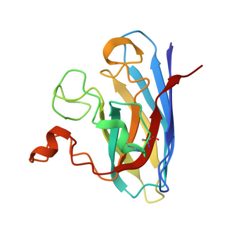
Ligand Binding and Aggregation of Pathogenic Sod1.

Wright, G.S.A.Antonyuk, S.V.Kershaw, N.M.Strange, R.W.Hasnain, S.S.

(2013) Nat Commun 4: 1758

  • DOI: https://doi.org/10.1038/ncomms2750
  • Primary Citation Related Structures: 
    4A7S, 4A7T, 4A7U, 4A7V

  • PubMed Abstract: 

    Mutations in the gene encoding Cu/Zn superoxide dismutase-1 cause amyotrophic lateral sclerosis. Superoxide dismutase-1 mutations decrease protein stability and promote aggregation. The mutant monomer is thought to be an intermediate in the pathway from the superoxide dismutase-1 dimer to aggregate. Here we find that the monomeric copper-apo, zinc-holo protein is structurally perturbed and the apo-protein aggregates without reattainment of the monomer-dimer equilibrium. Intervention to stabilize the superoxide dismutase-1 dimer and inhibit aggregation is regarded as a potential therapeutic strategy. We describe protein-ligand interactions for two compounds, Isoproterenol and 5-fluorouridine, highlighted as superoxide dismutase-1 stabilizers. We find both compounds interact with superoxide dismutase-1 at a key region identified at the core of the superoxide dismutase-1 fibrillar aggregates, β-barrel loop II-strand 3, rather than the proposed dimer interface site. This illustrates the need for direct structural observations when developing compounds for protein-targeted therapeutics.


  • Organizational Affiliation
    • Molecular Biophysics Group, Institute of Integrative Biology, Faculty of Health and Life Sciences, University of Liverpool, Liverpool L69 7ZB, UK.

Macromolecule Content 

  • Total Structure Weight: 32.43 kDa 
  • Atom Count: 2,592 
  • Modeled Residue Count: 306 
  • Deposited Residue Count: 306 
  • Unique protein chains: 1

Macromolecules

Find similar proteins by:|  3D Structure
Entity ID: 1
MoleculeChains  Sequence LengthOrganismDetailsImage
SUPEROXIDE DISMUTASE [CU-ZN]A,
B [auth F]
153Homo sapiensMutation(s): 1 
EC: 1.15.1.1 (PDB Primary Data), 1.8 (UniProt)
UniProt & NIH Common Fund Data Resources
Find proteins for P00441 (Homo sapiens)
Explore P00441 
Go to UniProtKB:  P00441
PHAROS:  P00441
GTEx:  ENSG00000142168 
Entity Groups
Sequence Clusters30% Identity50% Identity70% Identity90% Identity95% Identity100% Identity
UniProt GroupP00441
Sequence Annotations
Expand
Reference Sequence

Small Molecules

Ligands 4 Unique
IDChains Name / Formula / InChI Key2D Diagram3D Interactions
LDP

Query on LDP


Download:Ideal Coordinates CCD File
C [auth A]L-DOPAMINE
C8 H11 N O2
VYFYYTLLBUKUHU-UHFFFAOYSA-N
SO4

Query on SO4


Download:Ideal Coordinates CCD File
D [auth A],
E [auth A],
H [auth F],
I [auth F]
SULFATE ION
O4 S
QAOWNCQODCNURD-UHFFFAOYSA-L
ZN

Query on ZN


Download:Ideal Coordinates CCD File
G [auth A],
K [auth F]
ZINC ION
Zn
PTFCDOFLOPIGGS-UHFFFAOYSA-N
CU

Query on CU


Download:Ideal Coordinates CCD File
F [auth A],
J [auth F]
COPPER (II) ION
Cu
JPVYNHNXODAKFH-UHFFFAOYSA-N

Experimental Data & Validation

Experimental Data

  • Method: X-RAY DIFFRACTION
  • Resolution: 1.00 Å
  • R-Value Free:  0.181 (Depositor), 0.183 (DCC) 
  • R-Value Work:  0.158 (Depositor), 0.161 (DCC) 
  • R-Value Observed: 0.159 (Depositor) 
Space Group: P 1 21 1
Unit Cell:
Length ( Å )Angle ( ˚ )
a = 38.396α = 90
b = 68.035β = 104.82
c = 49.851γ = 90
Software Package:
Software NamePurpose
REFMACrefinement
HKL-2000data reduction
HKL-2000data scaling
REFMACphasing

Structure Validation

View Full Validation Report


Ligand Structure Quality Assessment 


Entry History 

Deposition Data

Revision History  (Full details and data files)

  • Version 1.0: 2012-11-28
    Type: Initial release
  • Version 1.1: 2013-04-24
    Changes: Database references
  • Version 1.2: 2013-05-08
    Changes: Database references
  • Version 1.3: 2024-10-23
    Changes: Data collection, Database references, Derived calculations, Other, Structure summary