4ZWZ | pdb_00004zwz

Engineered Carbonic Anhydrase IX mimic in complex with a glucosyl sulfamate inhibitor


Experimental Data Snapshot

  • Method: X-RAY DIFFRACTION
  • Resolution: 1.62 Å
  • R-Value Free: 
    0.211 (Depositor), 0.211 (DCC) 
  • R-Value Work: 
    0.161 (Depositor), 0.161 (DCC) 
  • R-Value Observed: 
    0.163 (Depositor) 

Starting Model: experimental
View more details

wwPDB Validation 3D Report Full Report

Validation slider image for 4ZWZ

Ligand Structure Quality Assessment 


This is version 1.3 of the entry. See complete history

Literature

Mapping Selective Inhibition of the Cancer-Related Carbonic Anhydrase IX Using Structure-Activity Relationships of Glucosyl-Based Sulfamates.

Mahon, B.P.Lomelino, C.L.Ladwig, J.Rankin, G.M.Driscoll, J.M.Salguero, A.L.Pinard, M.A.Vullo, D.Supuran, C.T.Poulsen, S.A.McKenna, R.

(2015) J Med Chem 58: 6630-6638

  • DOI: https://doi.org/10.1021/acs.jmedchem.5b00845
  • Primary Citation Related Structures: 
    4ZWX, 4ZWY, 4ZWZ, 4ZX0, 4ZX1

  • PubMed Abstract: 

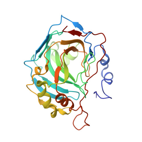
    Inhibition of human carbonic anhydrase IX (hCA IX) has shown to be therapeutically advantageous for treating many types of highly aggressive cancers. However, designing selective inhibitors for hCA IX has been difficult due to its high structural homology and sequence similarity with off-target hCAs. Recently, the use of glucosyl sulfamate inhibitors has shown promise as selective inhibitors for hCA IX. In this study, we present five X-ray crystal structures, determined to a resolution of 1.7 Å or better, of both hCA II (a ubiquitous CA) and an engineered hCA IX-mimic in complex with selected glucosyl sulfamates and structurally rationalize mechanisms for hCA IX selectivity. Results from this study have allowed us, for the first time, to empirically "map" key interactions of the hCA IX active site in order to establish parameters needed to design novel hCA IX selective inhibitors.


  • Organizational Affiliation
    • Department of Biochemistry and Molecular Biology, College of Medicine, University of Florida , 1600 SW Archer Road, PO Box 100245, Gainesville, Florida 32610, United States.

Macromolecule Content 

  • Total Structure Weight: 29.52 kDa 
  • Atom Count: 2,351 
  • Modeled Residue Count: 257 
  • Deposited Residue Count: 257 
  • Unique protein chains: 1

Macromolecules

Find similar proteins by:|  3D Structure
Entity ID: 1
MoleculeChains  Sequence LengthOrganismDetailsImage
Carbonic anhydrase 2257Homo sapiensMutation(s): 7 
Gene Names: CA2
EC: 4.2.1.1 (PDB Primary Data), 4.2.1.69 (UniProt)
UniProt & NIH Common Fund Data Resources
Find proteins for P00918 (Homo sapiens)
Explore P00918 
Go to UniProtKB:  P00918
PHAROS:  P00918
GTEx:  ENSG00000104267 
Entity Groups
Sequence Clusters30% Identity50% Identity70% Identity90% Identity95% Identity100% Identity
UniProt GroupP00918
Sequence Annotations
Expand
Reference Sequence

Experimental Data & Validation

Experimental Data

  • Method: X-RAY DIFFRACTION
  • Resolution: 1.62 Å
  • R-Value Free:  0.211 (Depositor), 0.211 (DCC) 
  • R-Value Work:  0.161 (Depositor), 0.161 (DCC) 
  • R-Value Observed: 0.163 (Depositor) 
Space Group: P 1 21 1
Unit Cell:
Length ( Å )Angle ( ˚ )
a = 41.695α = 90
b = 41.096β = 103.74
c = 71.43γ = 90
Software Package:
Software NamePurpose
HKL-2000data collection
HKL-2000data scaling
PHENIXrefinement
PDB_EXTRACTdata extraction
HKL-2000data reduction
PHENIXphasing

Structure Validation

View Full Validation Report


Ligand Structure Quality Assessment 


Entry History 

Deposition Data

Revision History  (Full details and data files)

  • Version 1.0: 2015-08-05
    Type: Initial release
  • Version 1.1: 2015-09-09
    Changes: Database references
  • Version 1.2: 2020-07-29
    Type: Remediation
    Reason: Carbohydrate remediation
    Changes: Database references, Derived calculations, Structure summary
  • Version 1.3: 2023-09-27
    Changes: Data collection, Database references, Refinement description