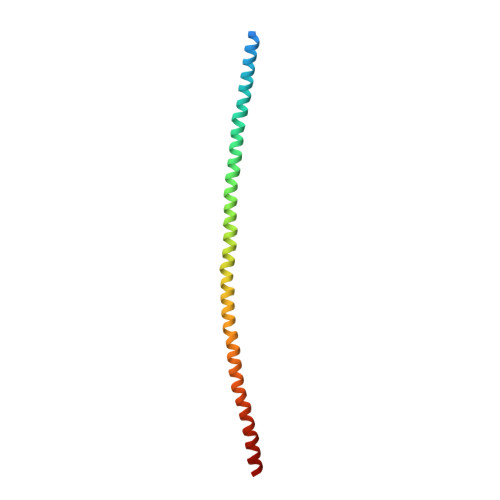
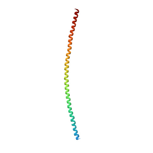
4ZRY

Crystal structure of the heterocomplex between coil 2B domains of human intermediate filament proteins keratin 1 (KRT1) and keratin 10 (KRT10)


Experimental Data Snapshot

  • Method: X-RAY DIFFRACTION
  • Resolution: 3.30 Å
  • R-Value Free: 0.277 
  • R-Value Work: 0.273 
  • R-Value Observed: 0.273 

Starting Model: experimental
View more details

wwPDB Validation   3D Report Full Report


This is version 1.3 of the entry. See complete history


Literature

The X-Ray Crystal Structure of the Keratin 1-Keratin 10 Helix 2B Heterodimer Reveals Molecular Surface Properties and Biochemical Insights into Human Skin Disease.

Bunick, C.G.Milstone, L.M.

(2017) J Invest Dermatol 137: 142-150

  • DOI: https://doi.org/10.1016/j.jid.2016.08.018
  • Primary Citation of Related Structures:  
    4ZRY

  • PubMed Abstract: 

    Keratins 1 (K1) and 10 (K10) are the primary keratins expressed in differentiated epidermis. Mutations in K1/K10 are associated with human skin diseases. We determined the crystal structure of the complex between the distal (2B) helices of K1 and K10 to better understand how human keratin structure correlates with function. The 3.3 Å resolution structure confirms many features inferred by previous biochemical analyses, but adds unexpected insights. It demonstrates a parallel, coiled-coil heterodimer with a predominantly hydrophobic intermolecular interface; this heterodimer formed a higher order complex with a second K1-K10-2B heterodimer via a Cys401 K10 disulfide link, although the bond angle is unanticipated. The molecular surface analysis of K1-K10-2B identified several pockets, one adjacent to the disulfide linkage and conserved in K5-K14. The solvent accessible surface area of the K1-K10 structure is 20-25% hydrophobic. The 2B region contains mixed acidic and basic patches proximally (N-terminal), whereas it is largely acidic distally (C-terminal). Mapping of conserved and nonconserved residues between K1-K10 and K5-K14 onto the structure demonstrated the majority of unique residues align along the outer helical ridge. Finally, the structure permitted a fresh analysis of the deleterious effects caused by K1/K10 missense mutations found in patients with phenotypic skin disease.


  • Organizational Affiliation

    Department of Dermatology, Yale University, New Haven, Connecticut, USA. Electronic address: christopher.bunick@yale.edu.


Macromolecules
Find similar proteins by:  (by identity cutoff)  |  3D Structure
Entity ID: 1
MoleculeChains Sequence LengthOrganismDetailsImage
Keratin, type I cytoskeletal 10124Homo sapiensMutation(s): 0 
Gene Names: KRT10KPP
UniProt & NIH Common Fund Data Resources
Find proteins for P13645 (Homo sapiens)
Explore P13645 
Go to UniProtKB:  P13645
PHAROS:  P13645
GTEx:  ENSG00000186395 
Entity Groups  
Sequence Clusters30% Identity50% Identity70% Identity90% Identity95% Identity100% Identity
UniProt GroupP13645
Sequence Annotations
Expand
  • Reference Sequence
Find similar proteins by:  (by identity cutoff)  |  3D Structure
Entity ID: 2
MoleculeChains Sequence LengthOrganismDetailsImage
Keratin, type II cytoskeletal 1120Homo sapiensMutation(s): 0 
Gene Names: KRT1KRTA
UniProt & NIH Common Fund Data Resources
Find proteins for P04264 (Homo sapiens)
Explore P04264 
Go to UniProtKB:  P04264
PHAROS:  P04264
GTEx:  ENSG00000167768 
Entity Groups  
Sequence Clusters30% Identity50% Identity70% Identity90% Identity95% Identity100% Identity
UniProt GroupP04264
Sequence Annotations
Expand
  • Reference Sequence
Experimental Data & Validation

Experimental Data

  • Method: X-RAY DIFFRACTION
  • Resolution: 3.30 Å
  • R-Value Free: 0.277 
  • R-Value Work: 0.273 
  • R-Value Observed: 0.273 
  • Space Group: I 2 2 2
Unit Cell:
Length ( Å )Angle ( ˚ )
a = 75.19α = 90
b = 75.86β = 90
c = 209.29γ = 90
Software Package:
Software NamePurpose
HKL-2000data collection
HKL-2000data scaling
XDSdata reduction
XDSdata scaling
MOLREPphasing
PHENIXrefinement
PDB_EXTRACTdata extraction

Structure Validation

View Full Validation Report


Entry History & Funding Information

Deposition Data


Funding OrganizationLocationGrant Number
Dermatology FoundationUnited States--

Revision History  (Full details and data files)

  • Version 1.0: 2016-05-18
    Type: Initial release
  • Version 1.1: 2017-01-11
    Changes: Database references
  • Version 1.2: 2017-11-22
    Changes: Refinement description
  • Version 1.3: 2023-09-27
    Changes: Data collection, Database references, Refinement description