A Versatile Strategy for the Semisynthetic Production of Ser65 Phosphorylated Ubiquitin and Its Biochemical and Structural Characterisation.
Han, C., Pao, K.C., Kazlauskaite, A., Muqit, M.M., Virdee, S.(2015) Chembiochem 16: 1574-1579
- PubMed: 26010437 Search on PubMedSearch on PubMed Central
- DOI: https://doi.org/10.1002/cbic.201500185
- Primary Citation Related Structures:
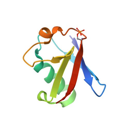
4ZPZ - PubMed Abstract:
Ubiquitin phosphorylation is emerging as an important regulatory layer in the ubiquitin system. This is exemplified by the phosphorylation of ubiquitin on Ser65 by the Parkinson's disease-associated kinase PINK1, which mediates the activation of the E3 ligase Parkin. Additional phosphorylation sites on ubiquitin might also have important cellular roles. Here we report a versatile strategy for preparing phosphorylated ubiquitin. We biochemically and structurally characterise semisynthetic phospho-Ser65-ubiquitin. Unexpectedly, we observed disulfide bond formation between ubiquitin molecules, and hence a novel crystal form. The method outlined provides a direct approach to study the combinatorial effects of phosphorylation on ubiquitin function. Our analysis also suggests that disulfide engineering of ubiquitin could be a useful strategy for obtaining alternative crystal forms of ubiquitin species thereby facilitating structural validation.
- MRC Protein Phosphorylation and Ubiquitylation Unit, College of Life Sciences, University of Dundee, Dow Street, Dundee DD1 5EH (UK).
Organizational Affiliation: