4ZMH | pdb_00004zmh

Crystal structure of a five-domain GH115 alpha-Glucuronidase from the Marine Bacterium Saccharophagus degradans 2-40T


Experimental Data Snapshot

  • Method: X-RAY DIFFRACTION
  • Resolution: 1.93 Å
  • R-Value Free: 
    0.170 (Depositor), 0.166 (DCC) 
  • R-Value Work: 
    0.138 (Depositor) 
  • R-Value Observed: 
    0.140 (Depositor) 

wwPDB Validation 3D Report Full Report

Validation slider image for 4ZMH

This is version 1.4 of the entry. See complete history

Literature

Biochemical and Structural Characterization of a Five-domain GH115 alpha-Glucuronidase from the Marine Bacterium Saccharophagus degradans 2-40T.

Wang, W.Yan, R.Nocek, B.P.Vuong, T.V.Di Leo, R.Xu, X.Cui, H.Gatenholm, P.Toriz, G.Tenkanen, M.Savchenko, A.Master, E.R.

(2016) J Biological Chem 291: 14120-14133

  • DOI: https://doi.org/10.1074/jbc.M115.702944
  • Primary Citation Related Structures: 
    4ZMH

  • PubMed Abstract: 

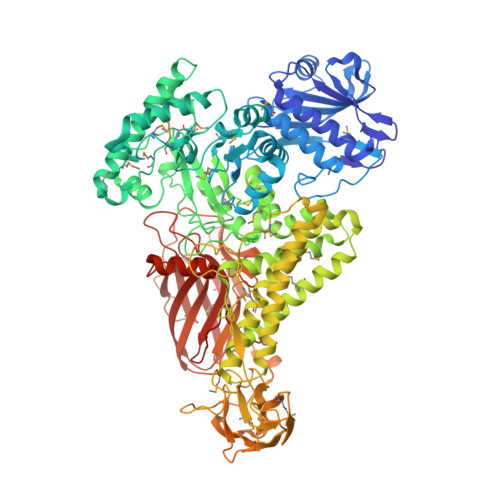
    Glucuronic acid (GlcAp) and/or methylglucuronic acid (MeGlcAp) decorate the major forms of xylan in hardwood and coniferous softwoods as well as many cereal grains. Accordingly, the complete utilization of glucuronoxylans or conversion to sugar precursors requires the action of main chain xylanases as well as α-glucuronidases that release the α- (1→2)-linked (Me)GlcAp side groups. Herein, a family GH115 enzymefrom the marine bacterium Saccharophagus degradans 2-40(T), SdeAgu115A, demonstrated activity toward glucuronoxylan and oligomers thereof with preference toward MeGlcAp linked to internal xylopyranosyl residues. Unique biochemical characteristics of NaCl activation were also observed. The crystal structure of SdeAgu115A revealed a five-domain architecture, with an additional insertion C(+) domain that had significant impact on the domain arrangement of SdeAgu115A monomer and its dimerization. The participation of domain C(+) in substrate binding was supported by reduced substrate inhibition upon introducing W773A, W689A, and F696A substitutions within this domain. In addition to Asp-335, the catalytic essentiality of Glu-216 was revealed by site-specific mutagenesis. A primary sequence analysis suggested that the SdeAgu115A architecture is shared by more than half of GH115 members, thus defining a distinct archetype for GH115 enzymes.


  • Organizational Affiliation
    • Department of Chemical Engineering and Applied Chemistry, University of Toronto, 200 College Street, Toronto, Ontario M5S 3E5, Canada.

Macromolecule Content 

  • Total Structure Weight: 214.98 kDa 
  • Atom Count: 17,210 
  • Modeled Residue Count: 1,866 
  • Deposited Residue Count: 1,878 
  • Unique protein chains: 1

Macromolecules

Find similar proteins by:|  3D Structure
Entity ID: 1
MoleculeChains  Sequence LengthOrganismDetailsImage
Uncharacterized protein
A, B
939Saccharophagus degradans 2-40Mutation(s): 0 
Gene Names: Sde_1755
UniProt
Find proteins for Q21JW4 (Saccharophagus degradans (strain 2-40 / ATCC 43961 / DSM 17024))
Explore Q21JW4 
Go to UniProtKB:  Q21JW4
Entity Groups
Sequence Clusters30% Identity50% Identity70% Identity90% Identity95% Identity100% Identity
UniProt GroupQ21JW4
Sequence Annotations
Expand
Reference Sequence

Small Molecules

Ligands 4 Unique
IDChains Name / Formula / InChI Key2D Diagram3D Interactions
PO4

Query on PO4


Download:Ideal Coordinates CCD File
C [auth A],
D [auth A],
E [auth A],
J [auth B]
PHOSPHATE ION
O4 P
NBIIXXVUZAFLBC-UHFFFAOYSA-K
GOL

Query on GOL


Download:Ideal Coordinates CCD File
G [auth A],
H [auth A]
GLYCEROL
C3 H8 O3
PEDCQBHIVMGVHV-UHFFFAOYSA-N
ACT

Query on ACT


Download:Ideal Coordinates CCD File
I [auth A]ACETATE ION
C2 H3 O2
QTBSBXVTEAMEQO-UHFFFAOYSA-M
NA

Query on NA


Download:Ideal Coordinates CCD File
F [auth A],
K [auth B]
SODIUM ION
Na
FKNQFGJONOIPTF-UHFFFAOYSA-N
Modified Residues  1 Unique
IDChains TypeFormula2D DiagramParent
MSE
Query on MSE
A, B
L-PEPTIDE LINKINGC5 H11 N O2 SeMET

Experimental Data & Validation

Experimental Data

  • Method: X-RAY DIFFRACTION
  • Resolution: 1.93 Å
  • R-Value Free:  0.170 (Depositor), 0.166 (DCC) 
  • R-Value Work:  0.138 (Depositor) 
  • R-Value Observed: 0.140 (Depositor) 
Space Group: P 21 21 21
Unit Cell:
Length ( Å )Angle ( ˚ )
a = 116.442α = 90
b = 124.294β = 90
c = 180.027γ = 90
Software Package:
Software NamePurpose
PHENIXrefinement
HKL-3000data scaling
HKL-3000phasing

Structure Validation

View Full Validation Report


Entry History 

Deposition Data

Revision History  (Full details and data files)

  • Version 1.0: 2016-05-04
    Type: Initial release
  • Version 1.1: 2016-05-11
    Changes: Database references
  • Version 1.2: 2016-05-18
    Changes: Structure summary
  • Version 1.3: 2016-07-13
    Changes: Database references
  • Version 1.4: 2024-11-06
    Changes: Data collection, Database references, Derived calculations, Structure summary