4ZH1

Complement factor H in complex with the GM1 glycan


Experimental Data Snapshot

  • Method: X-RAY DIFFRACTION
  • Resolution: 2.24 Å
  • R-Value Free: 0.228 
  • R-Value Work: 0.181 
  • R-Value Observed: 0.183 

wwPDB Validation   3D Report Full Report


This is version 2.1 of the entry. See complete history


Literature

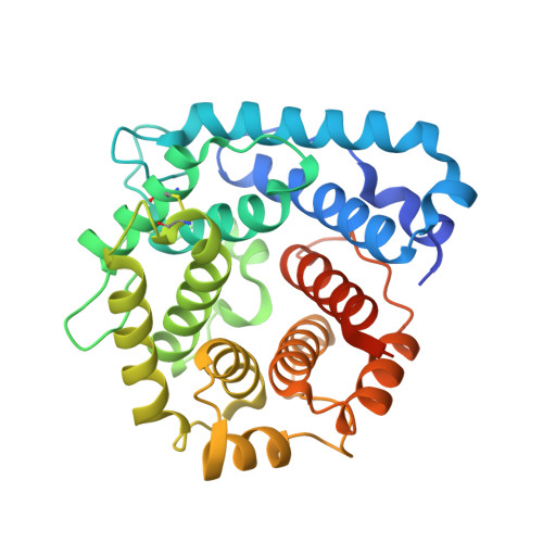
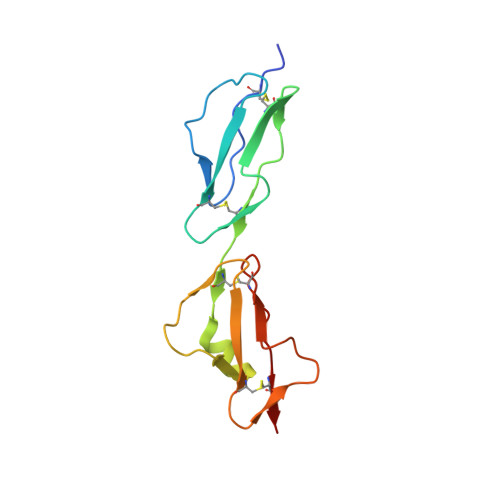
Complement Factor H and Simian Virus 40 bind the GM1 ganglioside in distinct conformations.

Blaum, B.S.Frank, M.Walker, R.C.Neu, U.Stehle, T.

(2016) Glycobiology 26: 532-539

  • DOI: https://doi.org/10.1093/glycob/cwv170
  • Primary Citation of Related Structures:  
    4ZH1

  • PubMed Abstract: 

    Mammalian cell surfaces are decorated with a variety of glycan chains that orchestrate development and defense and are exploited by pathogens for cellular attachment and entry. While glycosidic linkages are, in principle, flexible, the conformational space that a given glycan can sample is subject to spatial and electrostatic restrictions imposed by its overall chemical structure. Here, we show how the glycan moiety of the GM1 ganglioside, a branched, monosialylated pentasaccharide that serves as a ligand for various proteins, undergoes differential conformational selection in its interactions with different lectins. Using STD NMR and X-ray crystallography, we found that the innate immune regulator complement Factor H (FH) binds a previously not reported GM1 conformation that is not compatible with the GM1-binding sites of other structurally characterized GM1-binding lectins such as the Simian Virus 40 (SV40) capsid. Molecular dynamics simulations of the free glycan in explicit solvent on the 10 μs timescale reveal that the FH-bound conformation nevertheless corresponds to a minimum in the Gibbs free energy plot. In contrast to the GM1 conformation recognized by SV40, the FH-bound GM1 conformation is associated with poor NOE restraints, explaining how it escaped(1)H-(1)H NOE-restrained modeling in the past and highlighting the necessity for ensemble representations of glycan structures.


  • Organizational Affiliation

    Interfaculty Institute of Biochemistry, University of Tübingen, Tübingen 72076, Germany baerbel.blaum@uni-tuebingen.de.


Macromolecules
Find similar proteins by:  (by identity cutoff)  |  3D Structure
Entity ID: 1
MoleculeChains Sequence LengthOrganismDetailsImage
Complement C3
A, B, C
317Homo sapiensMutation(s): 0 
Gene Names: C3CPAMD1
UniProt & NIH Common Fund Data Resources
Find proteins for P01024 (Homo sapiens)
Explore P01024 
Go to UniProtKB:  P01024
PHAROS:  P01024
GTEx:  ENSG00000125730 
Entity Groups  
Sequence Clusters30% Identity50% Identity70% Identity90% Identity95% Identity100% Identity
UniProt GroupP01024
Sequence Annotations
Expand
  • Reference Sequence
Find similar proteins by:  (by identity cutoff)  |  3D Structure
Entity ID: 2
MoleculeChains Sequence LengthOrganismDetailsImage
Complement factor H
D, E, F
129Homo sapiensMutation(s): 0 
Gene Names: CFHHFHF1HF2
UniProt & NIH Common Fund Data Resources
Find proteins for P08603 (Homo sapiens)
Explore P08603 
Go to UniProtKB:  P08603
PHAROS:  P08603
GTEx:  ENSG00000000971 
Entity Groups  
Sequence Clusters30% Identity50% Identity70% Identity90% Identity95% Identity100% Identity
UniProt GroupP08603
Sequence Annotations
Expand
  • Reference Sequence
Oligosaccharides

Help

Entity ID: 3
MoleculeChains Length2D Diagram Glycosylation3D Interactions
N-acetyl-alpha-neuraminic acid-(2-3)-beta-D-galactopyranose
G
2N/A
Glycosylation Resources
GlyTouCan:  G30207PZ
GlyCosmos:  G30207PZ
GlyGen:  G30207PZ
Entity ID: 4
MoleculeChains Length2D Diagram Glycosylation3D Interactions
beta-D-galactopyranose-(1-3)-2-acetamido-2-deoxy-beta-D-galactopyranose-(1-4)-[N-acetyl-alpha-neuraminic acid-(2-3)]beta-D-galactopyranose-(1-4)-beta-D-glucopyranose
H
5N/A
Glycosylation Resources
GlyTouCan:  G48558GR
GlyCosmos:  G48558GR
GlyGen:  G48558GR
Entity ID: 5
MoleculeChains Length2D Diagram Glycosylation3D Interactions
N-acetyl-alpha-neuraminic acid-(2-3)-[2-acetamido-2-deoxy-beta-D-galactopyranose-(1-4)]beta-D-galactopyranose-(1-4)-beta-D-glucopyranose
I
4N/A
Glycosylation Resources
GlyTouCan:  G79389NT
GlyCosmos:  G79389NT
GlyGen:  G79389NT
Experimental Data & Validation

Experimental Data

  • Method: X-RAY DIFFRACTION
  • Resolution: 2.24 Å
  • R-Value Free: 0.228 
  • R-Value Work: 0.181 
  • R-Value Observed: 0.183 
  • Space Group: P 1
Unit Cell:
Length ( Å )Angle ( ˚ )
a = 75.77α = 112.97
b = 82.86β = 111.43
c = 86.57γ = 99.63
Software Package:
Software NamePurpose
REFMACrefinement
XDSdata reduction
Cootmodel building
XSCALEdata scaling
PHASERphasing

Structure Validation

View Full Validation Report


Entry History & Funding Information

Deposition Data


Funding OrganizationLocationGrant Number
German Research FoundationGermanyBL 1294/2-1

Revision History  (Full details and data files)

  • Version 1.0: 2016-01-13
    Type: Initial release
  • Version 1.1: 2016-01-20
    Changes: Structure summary
  • Version 1.2: 2016-04-13
    Changes: Database references
  • Version 2.0: 2020-07-29
    Type: Remediation
    Reason: Carbohydrate remediation
    Changes: Atomic model, Author supporting evidence, Data collection, Derived calculations, Structure summary
  • Version 2.1: 2024-01-10
    Changes: Data collection, Database references, Derived calculations, Refinement description, Structure summary