Oxaliplatin vs. cisplatin: competition experiments on their binding to lysozyme.
Marasco, D., Messori, L., Marzo, T., Merlino, A.(2015) Dalton Trans 44: 10392-10398
- PubMed: 25974859 Search on PubMed
- DOI: https://doi.org/10.1039/c5dt01279a
- Primary Citation Related Structures:
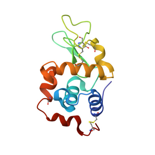
4Z46, 4ZEE - PubMed Abstract:
The model protein hen egg white lysozyme was challenged with oxaliplatin and cisplatin. ESI mass spectrometry, surface plasmon resonance and thermal shift analyses demonstrate the formation of a bis-platinum adduct, though in very small amounts. Crystals of the bis-platinum adduct were obtained using two different preparations and the X-ray structures were solved at 1.85 Å and 1.95 Å resolution. Overall, the obtained data point out that, under the analyzed conditions, the two Pt drugs have similar affinities for the protein, but bind on its surface at two non-overlapping sites. In other words, these two drugs manifest a significantly different reactivity with this model protein and do not compete for the same protein binding sites.
- Department of Pharmacy, University of Naples Federico II, via Montesano 12, 80120, Napoli, Italy.
Organizational Affiliation: