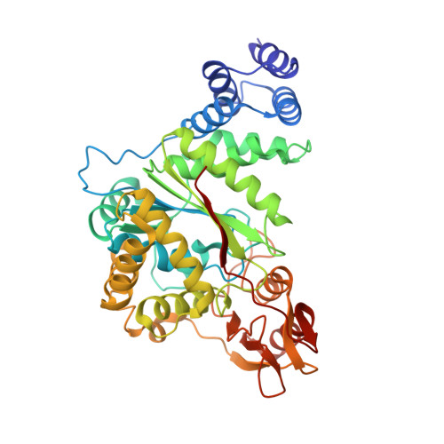
Structural investigation of the thymidine phosphorylase from Salmonella typhimurium in the unliganded state and its complexes with thymidine and uridine.
Balaev, V.V., Lashkov, A.A., Gabdulkhakov, A.G., Dontsova, M.V., Seregina, T.A., Mironov, A.S., Betzel, C., Mikhailov, A.M.(2016) Acta Crystallogr F Struct Biol Commun 72: 224-233
- PubMed: 26919527 Search on PubMedSearch on PubMed Central
- DOI: https://doi.org/10.1107/S2053230X1600162X
- Primary Citation Related Structures:
4XR5, 4YEK, 4YYY - PubMed Abstract:
Highly specific thymidine phosphorylases catalyze the phosphorolytic cleavage of thymidine, with the help of a phosphate ion, resulting in thymine and 2-deoxy-α-D-ribose 1-phosphate. Thymidine phosphorylases do not catalyze the phosphorolysis of uridine, in contrast to nonspecific pyrimidine nucleoside phosphorylases and uridine phosphorylases. Understanding the mechanism of substrate specificity on the basis of the nucleoside is essential to support rational drug-discovery investigations of new antitumour and anti-infective drugs which are metabolized by thymidine phosphorylases. For this reason, X-ray structures of the thymidine phosphorylase from Salmonella typhimurium were solved and refined: the unliganded structure at 2.05 Å resolution (PDB entry 4xr5), the structure of the complex with thymidine at 2.55 Å resolution (PDB entry 4yek) and that of the complex with uridine at 2.43 Å resolution (PDB entry 4yyy). The various structural features of the enzyme which might be responsible for the specificity for thymidine and not for uridine were identified. The presence of the 2'-hydroxyl group in uridine results in a different position of the uridine furanose moiety compared with that of thymidine. This feature may be the key element of the substrate specificity. The specificity might also be associated with the opening/closure mechanism of the two-domain subunit structure of the enzyme.
- A. V. Shubnikov Institute of Crystallography, Leninsky Prospect 59, Moscow 119333, Russian Federation.
Organizational Affiliation: