


Crystal structure of a RNA binding motif protein 39 (RBM39) from Homo sapiens at 1.28 A resolution
Joint Center for Structural Genomics (JCSG), Partnership for T-Cell Biology (TCELL)To be published.
Experimental Data Snapshot
Starting Model: experimental
View more details
wwPDB Validation 3D Report Full Report

Entity ID: 1 | |||||
|---|---|---|---|---|---|
| Molecule | Chains | Sequence Length | Organism | Details | Image |
| RNA-binding protein 39 | 92 | Homo sapiens | Mutation(s): 0 Gene Names: RBM39, HCC1, RNPC2 | 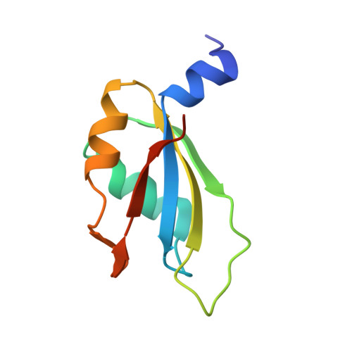 | |
UniProt & NIH Common Fund Data Resources | |||||
PHAROS: Q14498 GTEx: ENSG00000131051 | |||||
Entity Groups | |||||
| Sequence Clusters | 30% Identity50% Identity70% Identity90% Identity95% Identity100% Identity | ||||
| UniProt Group | Q14498 | ||||
Sequence AnnotationsExpand | |||||
Reference Sequence | |||||
| Length ( Å ) | Angle ( ˚ ) |
|---|---|
| a = 86.574 | α = 90 |
| b = 26.711 | β = 90 |
| c = 35.015 | γ = 90 |
| Software Name | Purpose |
|---|---|
| PDB_EXTRACT | data extraction |
| PHASER | phasing |
| REFMAC | refinement |
| XDS | data reduction |
| XSCALE | data scaling |
| SCALA | data scaling |


