4YH2

Glutathione Transferase E6 from Drosophila melanogaster


Experimental Data Snapshot

  • Method: X-RAY DIFFRACTION
  • Resolution: 1.72 Å
  • R-Value Free: 0.188 
  • R-Value Work: 0.157 
  • R-Value Observed: 0.159 

wwPDB Validation   3D Report Full Report


Ligand Structure Quality Assessment 


This is version 1.2 of the entry. See complete history


Literature

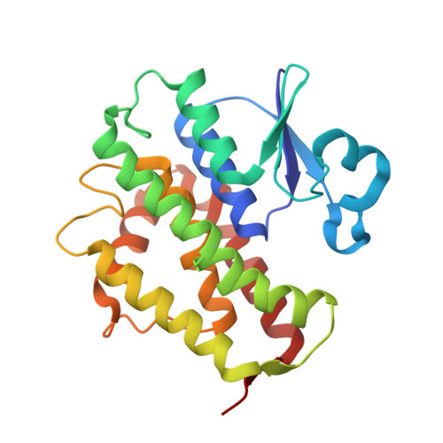
Epsilon glutathione transferases possess a unique class-conserved subunit interface motif that directly interacts with glutathione in the active site

Wongsantichon, J.Robinson, R.C.Ketterman, A.J.

(2015) Biosci Rep 35

  • DOI: https://doi.org/10.1042/BSR20150183
  • Primary Citation of Related Structures:  
    4YH2

  • PubMed Abstract: 

    Epsilon class glutathione transferases (GSTs) have been shown to contribute significantly to insecticide resistance. We report a new Epsilon class protein crystal structure from Drosophila melanogaster for the glutathione transferase DmGSTE6. The structure reveals a novel Epsilon clasp motif that is conserved across hundreds of millions of years of evolution of the insect Diptera order. This histidine-serine motif lies in the subunit interface and appears to contribute to quaternary stability as well as directly connecting the two glutathiones in the active sites of this dimeric enzyme.


  • Organizational Affiliation

    Institute of Molecular and Cell Biology, A*STAR (Agency for Science, Technology and Research), Biopolis, Singapore 138673.


Macromolecules
Find similar proteins by:  (by identity cutoff)  |  3D Structure
Entity ID: 1
MoleculeChains Sequence LengthOrganismDetailsImage
Glutathione S transferase E6
A, B, C, D
222Drosophila melanogasterMutation(s): 0 
Gene Names: GstE6CG17530Dmel_CG17530
EC: 2.5.1.18
UniProt
Find proteins for A1ZB71 (Drosophila melanogaster)
Explore A1ZB71 
Go to UniProtKB:  A1ZB71
Entity Groups  
Sequence Clusters30% Identity50% Identity70% Identity90% Identity95% Identity100% Identity
UniProt GroupA1ZB71
Sequence Annotations
Expand
  • Reference Sequence
Experimental Data & Validation

Experimental Data

  • Method: X-RAY DIFFRACTION
  • Resolution: 1.72 Å
  • R-Value Free: 0.188 
  • R-Value Work: 0.157 
  • R-Value Observed: 0.159 
  • Space Group: C 1 2 1
Unit Cell:
Length ( Å )Angle ( ˚ )
a = 176.35α = 90
b = 58.86β = 128.25
c = 122.81γ = 90
Software Package:
Software NamePurpose
Aimlessdata scaling
PHENIXrefinement
PDB_EXTRACTdata extraction
xia2data reduction
xia2phasing

Structure Validation

View Full Validation Report


Ligand Structure Quality Assessment 


Entry History 

Deposition Data

Revision History  (Full details and data files)

  • Version 1.0: 2016-02-10
    Type: Initial release
  • Version 1.1: 2020-02-05
    Changes: Data collection, Database references, Derived calculations
  • Version 1.2: 2024-03-20
    Changes: Data collection, Database references