4YH1 | pdb_00004yh1

Structure of Human Scp1 bound to cis-proline peptidomimetic CTD phospho-Ser5 peptide


Experimental Data Snapshot

  • Method: X-RAY DIFFRACTION
  • Resolution: 2.20 Å
  • R-Value Free: 
    0.246 (Depositor), 0.254 (DCC) 
  • R-Value Work: 
    0.193 (Depositor), 0.199 (DCC) 
  • R-Value Observed: 
    0.195 (Depositor) 

wwPDB Validation 3D Report Full Report

Validation slider image for 4YH1

This is version 1.2 of the entry. See complete history

Literature

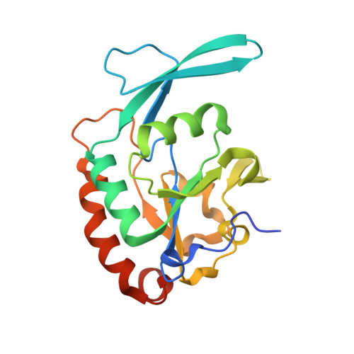
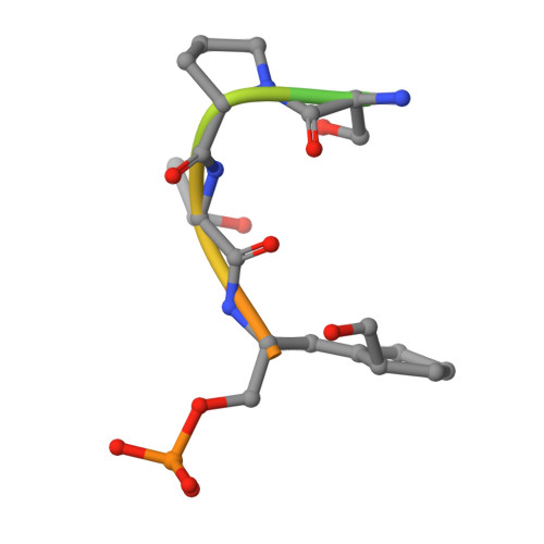
Chemical Tools To Decipher Regulation of Phosphatases by Proline Isomerization on Eukaryotic RNA Polymerase II.

Mayfield, J.E.Fan, S.Wei, S.Zhang, M.Li, B.Ellington, A.D.Etzkorn, F.A.Zhang, Y.J.

(2015) ACS Chem Biol 10: 2405-2414

  • DOI: https://doi.org/10.1021/acschembio.5b00296
  • Primary Citation Related Structures: 
    4YGX, 4YGY, 4YH1

  • PubMed Abstract: 

    Proline isomerization greatly impacts biological signaling but is subtle and difficult to detect in proteins. We characterize this poorly understood regulatory mechanism for RNA polymerase II carboxyl terminal domain (CTD) phosphorylation state using novel, direct, and quantitative chemical tools. We determine the proline isomeric preference of three CTD phosphatases: Ssu72 as cis-proline specific, Scp1 and Fcp1 as strongly trans-preferred. Due to this inherent characteristic, these phosphatases respond differently to enzymes that catalyze the isomerization of proline, like Ess1/Pin1. We demonstrate that this selective regulation of RNA polymerase II phosphorylation state exists within human cells, consistent with in vitro assays. These results support a model in which, instead of a global enhancement of downstream enzymatic activities, proline isomerases selectively boost the activity of a subset of CTD regulatory factors specific for cis-proline. This leads to diversified phosphorylation states of CTD in vitro and in cells. We provide the chemical tools to investigate proline isomerization and its ability to selectively enhance signaling in transcription and other biological contexts.


  • Organizational Affiliation
    • Department of Molecular Biosciences and Institute for Cellular and Molecular Biology, University of Texas at Austin , Austin, Texas 78712, United States.

Macromolecule Content 

  • Total Structure Weight: 45.71 kDa 
  • Atom Count: 3,123 
  • Modeled Residue Count: 366 
  • Deposited Residue Count: 402 
  • Unique protein chains: 2

Macromolecules

Find similar proteins by:|  3D Structure
Entity ID: 1
MoleculeChains  Sequence LengthOrganismDetailsImage
Carboxy-terminal domain RNA polymerase II polypeptide A small phosphatase 1
A, B
189Homo sapiensMutation(s): 1 
Gene Names: CTDSP1NIF3NLIIFSCP1
EC: 3.1.3.16
UniProt & NIH Common Fund Data Resources
Find proteins for Q9GZU7 (Homo sapiens)
Explore Q9GZU7 
Go to UniProtKB:  Q9GZU7
PHAROS:  Q9GZU7
GTEx:  ENSG00000144579 
Entity Groups
Sequence Clusters30% Identity50% Identity70% Identity90% Identity95% Identity100% Identity
UniProt GroupQ9GZU7
Sequence Annotations
Expand
Reference Sequence
Find similar proteins by:  Sequence   |   3D Structure  
Entity ID: 2
MoleculeChains  Sequence LengthOrganismDetailsImage
A small phosphatase 1
C, D
12synthetic constructMutation(s): 0 

Experimental Data & Validation

Experimental Data

  • Method: X-RAY DIFFRACTION
  • Resolution: 2.20 Å
  • R-Value Free:  0.246 (Depositor), 0.254 (DCC) 
  • R-Value Work:  0.193 (Depositor), 0.199 (DCC) 
  • R-Value Observed: 0.195 (Depositor) 
Space Group: C 1 2 1
Unit Cell:
Length ( Å )Angle ( ˚ )
a = 125.115α = 90
b = 78.804β = 112.54
c = 62.853γ = 90
Software Package:
Software NamePurpose
HKL-2000data reduction
PHASERphasing
REFMACrefinement
PDB_EXTRACTdata extraction

Structure Validation

View Full Validation Report


Entry History 

Deposition Data

Revision History  (Full details and data files)

  • Version 1.0: 2015-09-16
    Type: Initial release
  • Version 1.1: 2015-10-28
    Changes: Database references
  • Version 1.2: 2024-10-30
    Changes: Data collection, Database references, Derived calculations, Structure summary