Crystallography and chemistry should always go together: a cautionary tale of protein complexes with cisplatin and carboplatin.
Shabalin, I., Dauter, Z., Jaskolski, M., Minor, W., Wlodawer, A.(2015) Acta Crystallogr D Biol Crystallogr 71: 1965-1979
- PubMed: 26327386 Search on PubMedSearch on PubMed Central
- DOI: https://doi.org/10.1107/S139900471500629X
- Primary Citation Related Structures:
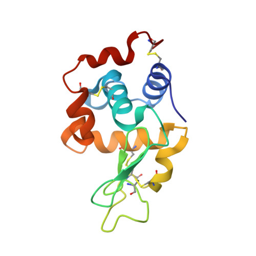
4YDX, 4YEA, 4YEM, 4YEN, 4YEO - PubMed Abstract:
The anticancer activity of platinum-containing drugs such as cisplatin and carboplatin is considered to primarily arise from their interactions with nucleic acids; nevertheless, these drugs, or the products of their hydrolysis, also bind to proteins, potentially leading to the known side effects of the treatments. Here, over 40 crystal structures deposited in the Protein Data Bank (PDB) of cisplatin and carboplatin complexes of several proteins were analysed. Significant problems of either a crystallographic or a chemical nature were found in most of the presented atomic models and they could be traced to less or more serious deficiencies in the data-collection and refinement procedures. The re-evaluation of these data and models was possible thanks to their mandatory or voluntary deposition in publicly available databases, emphasizing the point that the availability of such data is critical for making structural science reproducible. Based on this analysis of a selected group of macromolecular structures, the importance of deposition of raw diffraction data is stressed and a procedure for depositing, tracking and using re-refined crystallographic models is suggested.
- Department of Molecular Physiology and Biological Physics, University of Virginia, 1340 Jefferson Park Avenue, Charlottesville, VA 22908, USA.
Organizational Affiliation: