4Y8W

Crystal Structure of Human Cytochrome P450 21A2 Progesterone Complex


Experimental Data Snapshot

  • Method: X-RAY DIFFRACTION
  • Resolution: 2.64 Å
  • R-Value Free: 0.281 
  • R-Value Work: 0.231 
  • R-Value Observed: 0.235 

wwPDB Validation   3D Report Full Report


Ligand Structure Quality Assessment 


This is version 1.5 of the entry. See complete history


Literature

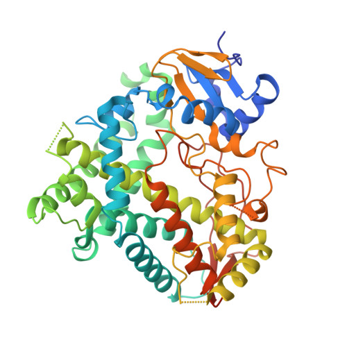
Human Cytochrome P450 21A2, the Major Steroid 21-Hydroxylase: STRUCTURE OF THE ENZYMEPROGESTERONE SUBSTRATE COMPLEX AND RATE-LIMITING C-H BOND CLEAVAGE.

Pallan, P.S.Wang, C.Lei, L.Yoshimoto, F.K.Auchus, R.J.Waterman, M.R.Guengerich, F.P.Egli, M.

(2015) J Biol Chem 290: 13128-13143

  • DOI: https://doi.org/10.1074/jbc.M115.646307
  • Primary Citation of Related Structures:  
    4Y8W

  • PubMed Abstract: 

    Cytochrome P450 (P450) 21A2 is the major steroid 21-hydroxylase, and deficiency of this enzyme is involved in ∼95% of cases of human congenital adrenal hyperplasia, a disorder of adrenal steroidogenesis. A structure of the bovine enzyme that we published previously (Zhao, B., Lei, L., Kagawa, N., Sundaramoorthy, M., Banerjee, S., Nagy, L. D., Guengerich, F. P., and Waterman, M. R. (2012) Three-dimensional structure of steroid 21-hydroxylase (cytochrome P450 21A2) with two substrates reveals locations of disease-associated variants. J. Biol. Chem. 287, 10613-10622), containing two molecules of the substrate 17α-hydroxyprogesterone, has been used as a template for understanding genetic deficiencies. We have now obtained a crystal structure of human P450 21A2 in complex with progesterone, a substrate in adrenal 21-hydroxylation. Substrate binding and release were fast for human P450 21A2 with both substrates, and pre-steady-state kinetics showed a partial burst but only with progesterone as substrate and not 17α-hydroxyprogesterone. High intermolecular non-competitive kinetic deuterium isotope effects on both kcat and kcat/Km, from 5 to 11, were observed with both substrates, indicative of rate-limiting C-H bond cleavage and suggesting that the juxtaposition of the C21 carbon in the active site is critical for efficient oxidation. The estimated rate of binding of the substrate progesterone (kon 2.4 × 10(7) M(-1) s(-1)) is only ∼2-fold greater than the catalytic efficiency (kcat/Km = 1.3 × 10(7) M(-1) s(-1)) with this substrate, suggesting that the rate of substrate binding may also be partially rate-limiting. The structure of the human P450 21A2-substrate complex provides direct insight into mechanistic effects of genetic variants.


  • Organizational Affiliation

    From the Department of Biochemistry, Vanderbilt University School of Medicine, Nashville, Tennessee 37232-0146 and.


Macromolecules
Find similar proteins by:  (by identity cutoff)  |  3D Structure
Entity ID: 1
MoleculeChains Sequence LengthOrganismDetailsImage
Cytochrome P450 21-hydroxylase
A, B, C
482Homo sapiensMutation(s): 0 
Gene Names: P450-CYP21BCYP21A2hCG_1999926
Membrane Entity: Yes 
UniProt & NIH Common Fund Data Resources
Find proteins for P08686 (Homo sapiens)
Explore P08686 
Go to UniProtKB:  P08686
PHAROS:  P08686
GTEx:  ENSG00000231852 
Entity Groups  
Sequence Clusters30% Identity50% Identity70% Identity90% Identity95% Identity100% Identity
UniProt GroupP08686
Sequence Annotations
Expand
  • Reference Sequence
Small Molecules
Ligands 3 Unique
IDChains Name / Formula / InChI Key2D Diagram3D Interactions
HEM
Query on HEM

Download Ideal Coordinates CCD File 
F [auth A],
I [auth B],
L [auth C]
PROTOPORPHYRIN IX CONTAINING FE
C34 H32 Fe N4 O4
KABFMIBPWCXCRK-RGGAHWMASA-L
STR
Query on STR

Download Ideal Coordinates CCD File 
G [auth A],
J [auth B],
M [auth C]
PROGESTERONE
C21 H30 O2
RJKFOVLPORLFTN-LEKSSAKUSA-N
SO4
Query on SO4

Download Ideal Coordinates CCD File 
D [auth A],
E [auth A],
H [auth B],
K [auth C]
SULFATE ION
O4 S
QAOWNCQODCNURD-UHFFFAOYSA-L
Experimental Data & Validation

Experimental Data

  • Method: X-RAY DIFFRACTION
  • Resolution: 2.64 Å
  • R-Value Free: 0.281 
  • R-Value Work: 0.231 
  • R-Value Observed: 0.235 
  • Space Group: C 1 2 1
Unit Cell:
Length ( Å )Angle ( ˚ )
a = 150.383α = 90
b = 86.86β = 102.11
c = 108.923γ = 90
Software Package:
Software NamePurpose
REFMACrefinement
HKL-2000data reduction
HKL-2000data scaling
MOLREPphasing

Structure Validation

View Full Validation Report


Ligand Structure Quality Assessment 


Entry History & Funding Information

Deposition Data


Funding OrganizationLocationGrant Number
National Institutes of Health/National Institute of General Medical Sciences (NIH/NIGMS)United StatesGM103937

Revision History  (Full details and data files)

  • Version 1.0: 2015-04-15
    Type: Initial release
  • Version 1.1: 2015-04-22
    Changes: Database references
  • Version 1.2: 2015-06-03
    Changes: Database references
  • Version 1.3: 2017-09-20
    Changes: Advisory, Author supporting evidence, Database references, Derived calculations, Source and taxonomy
  • Version 1.4: 2019-12-25
    Changes: Author supporting evidence
  • Version 1.5: 2023-09-27
    Changes: Data collection, Database references, Derived calculations, Refinement description