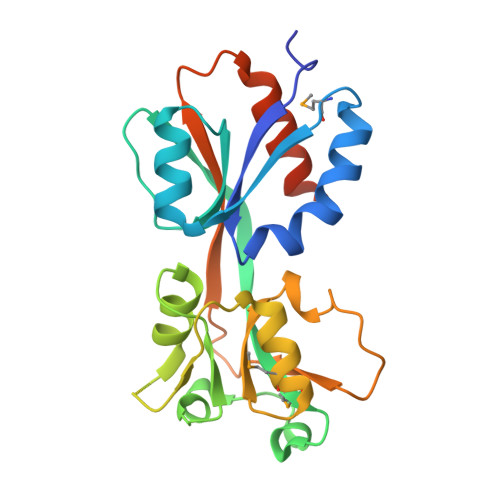
4Y0M

The reduced form of OxyR regulatory domain from Psedomonas aeruginosa


Experimental Data Snapshot

  • Method: X-RAY DIFFRACTION
  • Resolution: 2.30 Å
  • R-Value Free: 0.271 
  • R-Value Work: 0.217 
  • R-Value Observed: 0.219 

wwPDB Validation   3D Report Full Report


This is version 1.3 of the entry. See complete history


Literature

Structural details of the OxyR peroxide-sensing mechanism

Jo, I.Chung, I.Y.Bae, H.W.Kim, J.S.Song, S.Cho, Y.H.Ha, N.C.

(2015) Proc Natl Acad Sci U S A 112: 6443-6448

  • DOI: https://doi.org/10.1073/pnas.1424495112
  • Primary Citation of Related Structures:  
    4X6G, 4XWS, 4Y0M

  • PubMed Abstract: 

    OxyR, a bacterial peroxide sensor, is a LysR-type transcriptional regulator (LTTR) that regulates the transcription of defense genes in response to a low level of cellular H2O2. Consisting of an N-terminal DNA-binding domain (DBD) and a C-terminal regulatory domain (RD), OxyR senses H2O2 with conserved cysteine residues in the RD. However, the precise mechanism of OxyR is not yet known due to the absence of the full-length (FL) protein structure. Here we determined the crystal structures of the FL protein and RD of Pseudomonas aeruginosa OxyR and its C199D mutant proteins. The FL crystal structures revealed that OxyR has a tetrameric arrangement assembled via two distinct dimerization interfaces. The C199D mutant structures suggested that new interactions that are mediated by cysteine hydroxylation induce a large conformational change, facilitating intramolecular disulfide-bond formation. More importantly, a bound H2O2 molecule was found near the Cys199 site, suggesting the H2O2-driven oxidation mechanism of OxyR. Combined with the crystal structures, a modeling study suggested that a large movement of the DBD is triggered by structural changes in the regulatory domains upon oxidation. Taken together, these findings provide novel concepts for answering key questions regarding OxyR in the H2O2-sensing and oxidation-dependent regulation of antioxidant genes.


  • Organizational Affiliation

    Department of Agricultural Biotechnology, Center for Food Safety and Toxicology, Center for Food and Bioconvergence, Research Institute for Agricultural and Life Sciences, Seoul National University, Seoul 151-921, Republic of Korea; and.


Macromolecules
Find similar proteins by:  (by identity cutoff)  |  3D Structure
Entity ID: 1
MoleculeChains Sequence LengthOrganismDetailsImage
OxyR
A, B, C, D, E
A, B, C, D, E, F, G, H, I, J, K, L
227Pseudomonas aeruginosa PAO1Mutation(s): 0 
Gene Names: oxyRPA5344
UniProt
Find proteins for Q9HTL4 (Pseudomonas aeruginosa (strain ATCC 15692 / DSM 22644 / CIP 104116 / JCM 14847 / LMG 12228 / 1C / PRS 101 / PAO1))
Explore Q9HTL4 
Go to UniProtKB:  Q9HTL4
Entity Groups  
Sequence Clusters30% Identity50% Identity70% Identity90% Identity95% Identity100% Identity
UniProt GroupQ9HTL4
Sequence Annotations
Expand
  • Reference Sequence
Small Molecules
Modified Residues  1 Unique
IDChains TypeFormula2D DiagramParent
MSE
Query on MSE
A, B, C, D, E
A, B, C, D, E, F, G, H, I, J, K, L
L-PEPTIDE LINKINGC5 H11 N O2 SeMET
Experimental Data & Validation

Experimental Data

  • Method: X-RAY DIFFRACTION
  • Resolution: 2.30 Å
  • R-Value Free: 0.271 
  • R-Value Work: 0.217 
  • R-Value Observed: 0.219 
  • Space Group: P 31 1 2
Unit Cell:
Length ( Å )Angle ( ˚ )
a = 151.355α = 90
b = 151.355β = 90
c = 218.844γ = 120
Software Package:
Software NamePurpose
PHENIXrefinement
d*TREKdata reduction
HKL-2000data scaling
PHASERphasing

Structure Validation

View Full Validation Report


Entry History 

Deposition Data

Revision History  (Full details and data files)

  • Version 1.0: 2015-04-29
    Type: Initial release
  • Version 1.1: 2015-06-03
    Changes: Database references
  • Version 1.2: 2018-05-16
    Changes: Data collection, Database references, Derived calculations, Source and taxonomy, Structure summary
  • Version 1.3: 2024-11-20
    Changes: Data collection, Database references, Structure summary