4XZ2 | pdb_00004xz2

Human platelet phosphofructokinase in an R-state in complex with ADP and F6P, crystal form I


Experimental Data Snapshot

  • Method: X-RAY DIFFRACTION
  • Resolution: 2.67 Å
  • R-Value Free: 
    0.255 (Depositor), 0.255 (DCC) 
  • R-Value Work: 
    0.216 (Depositor), 0.219 (DCC) 
  • R-Value Observed: 
    0.218 (Depositor) 

Starting Model: experimental
View more details

wwPDB Validation 3D Report Full Report

Validation slider image for 4XZ2

Ligand Structure Quality Assessment 


This is version 1.4 of the entry. See complete history

Literature

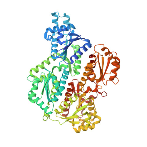
Crystal structure of human platelet phosphofructokinase-1 locked in an activated conformation.

Kloos, M.Bruser, A.Kirchberger, J.Schoneberg, T.Strater, N.

(2015) Biochem J 469: 421-432

  • DOI: https://doi.org/10.1042/BJ20150251
  • Primary Citation Related Structures: 
    4RH3, 4U1R, 4WL0, 4XZ2

  • PubMed Abstract: 

    Phosphofructokinase-1 (Pfk) acts as the main control point of flux through glycolysis. It is involved in complex allosteric regulation and Pfk mutations have been linked to cancer development. Whereas the 3D structure and structural basis of allosteric regulation of prokaryotic Pfk has been studied in great detail, our knowledge about the molecular basis of the allosteric behaviour of the more complex mammalian Pfk is still very limited. To characterize the structural basis of allosteric regulation, the subunit interfaces and the functional consequences of modifications in Tarui's disease and cancer, we analysed the physiological homotetramer of human platelet Pfk at up to 2.67 Å resolution in two crystal forms. The crystallized enzyme is permanently activated by a deletion of the 22 C-terminal residues. Complex structures with ADP and fructose-6-phosphate (F6P) and with ATP suggest a role of three aspartates in the deprotonation of the OH-nucleophile of F6P and in the co-ordination of the catalytic magnesium ion. Changes at the dimer interface, including an asymmetry observed in both crystal forms, are the primary mechanism of allosteric regulation of Pfk by influencing the F6P-binding site. Whereas the nature of this conformational switch appears to be largely conserved in bacterial, yeast and mammalian Pfk, initiation of these changes differs significantly in eukaryotic Pfk.


  • Organizational Affiliation
    • Institute of Bioanalytical Chemistry, Center for Biotechnology and Biomedicine, University of Leipzig, Deutscher Platz 5, 04103 Leipzig, Germany.

Macromolecule Content 

  • Total Structure Weight: 335.95 kDa 
  • Atom Count: 22,132 
  • Modeled Residue Count: 2,865 
  • Deposited Residue Count: 3,044 
  • Unique protein chains: 1

Macromolecules

Find similar proteins by:|  3D Structure
Entity ID: 1
MoleculeChains  Sequence LengthOrganismDetailsImage
ATP-dependent 6-phosphofructokinase, platelet type
A, B, C, D
761Homo sapiensMutation(s): 0 
Gene Names: PFKPPFKF
EC: 2.7.1.11
UniProt & NIH Common Fund Data Resources
Find proteins for Q01813 (Homo sapiens)
Explore Q01813 
Go to UniProtKB:  Q01813
PHAROS:  Q01813
GTEx:  ENSG00000067057 
Entity Groups
Sequence Clusters30% Identity50% Identity70% Identity90% Identity95% Identity100% Identity
UniProt GroupQ01813
Sequence Annotations
Expand
Reference Sequence

Small Molecules

Ligands 4 Unique
IDChains Name / Formula / InChI Key2D Diagram3D Interactions
ADP

Query on ADP


Download:Ideal Coordinates CCD File
G [auth B],
N [auth D]
ADENOSINE-5'-DIPHOSPHATE
C10 H15 N5 O10 P2
XTWYTFMLZFPYCI-KQYNXXCUSA-N
FBP

Query on FBP


Download:Ideal Coordinates CCD File
F [auth A],
H [auth B],
L [auth C],
O [auth D]
1,6-di-O-phosphono-beta-D-fructofuranose
C6 H14 O12 P2
RNBGYGVWRKECFJ-ARQDHWQXSA-N
F6P

Query on F6P


Download:Ideal Coordinates CCD File
I [auth B],
P [auth D]
6-O-phosphono-beta-D-fructofuranose
C6 H13 O9 P
BGWGXPAPYGQALX-ARQDHWQXSA-N
PO4

Query on PO4


Download:Ideal Coordinates CCD File
E [auth A]
J [auth B]
K [auth B]
M [auth C]
Q [auth D]
E [auth A],
J [auth B],
K [auth B],
M [auth C],
Q [auth D],
R [auth D]
PHOSPHATE ION
O4 P
NBIIXXVUZAFLBC-UHFFFAOYSA-K

Experimental Data & Validation

Experimental Data

  • Method: X-RAY DIFFRACTION
  • Resolution: 2.67 Å
  • R-Value Free:  0.255 (Depositor), 0.255 (DCC) 
  • R-Value Work:  0.216 (Depositor), 0.219 (DCC) 
  • R-Value Observed: 0.218 (Depositor) 
Space Group: P 1 21 1
Unit Cell:
Length ( Å )Angle ( ˚ )
a = 76.652α = 90
b = 164.524β = 102.96
c = 133.2γ = 90
Software Package:
Software NamePurpose
REFMACrefinement
XDSdata reduction
PHASERphasing
PDB_EXTRACTdata extraction
XSCALEdata scaling

Structure Validation

View Full Validation Report


Ligand Structure Quality Assessment 


Entry History 

& Funding Information

Deposition Data


Funding OrganizationLocationGrant Number
German Research FoundationGermanySFB 610

Revision History  (Full details and data files)

  • Version 1.0: 2015-06-03
    Type: Initial release
  • Version 1.1: 2015-07-01
    Changes: Database references
  • Version 1.2: 2015-08-05
    Changes: Database references
  • Version 1.3: 2020-07-29
    Type: Remediation
    Reason: Carbohydrate remediation
    Changes: Author supporting evidence, Data collection, Derived calculations, Structure summary
  • Version 1.4: 2024-01-10
    Changes: Data collection, Database references, Refinement description, Structure summary