4XLV | pdb_00004xlv

Crystal structure of the activated insulin receptor tyrosine kinase dimer

  • Classification: TRANSFERASE
  • Organism(s): Homo sapiens
  • Expression System: Spodoptera frugiperda
  • Mutation(s): Yes 

  • Deposited: 2015-01-13 Released: 2015-03-25 
  • Deposition Author(s): Hubbard, S.R., Li, S.
  • Funding Organization(s): National Institutes of Health/National Institute of Diabetes and Digestive and Kidney Disease (NIH/NIDDK)

Experimental Data Snapshot

  • Method: X-RAY DIFFRACTION
  • Resolution: 2.30 Å
  • R-Value Free: 
    0.266 (Depositor), 0.264 (DCC) 
  • R-Value Work: 
    0.204 (Depositor), 0.199 (DCC) 
  • R-Value Observed: 
    0.207 (Depositor) 

wwPDB Validation 3D Report Full Report

Validation slider image for 4XLV

Ligand Structure Quality Assessment 


This is version 1.3 of the entry. See complete history

Literature

The insulin and IGF1 receptor kinase domains are functional dimers in the activated state.

Cabail, M.Z.Li, S.Lemmon, E.Bowen, M.E.Hubbard, S.R.Miller, W.T.

(2015) Nat Commun 6: 6406-6406

  • DOI: https://doi.org/10.1038/ncomms7406
  • Primary Citation Related Structures: 
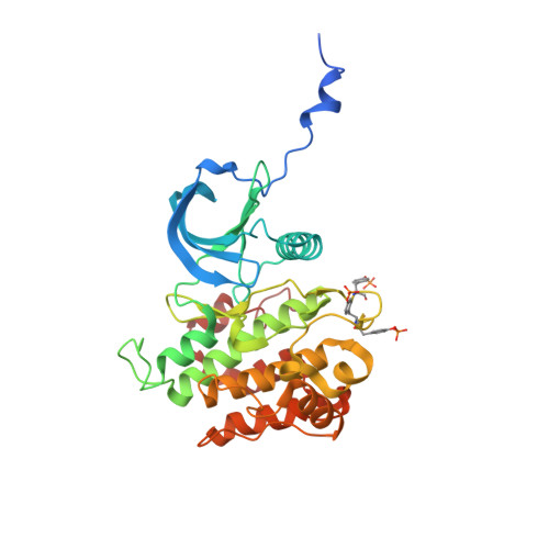
    4XLV

  • PubMed Abstract: 

    The insulin receptor (IR) and insulin-like growth factor-1 receptor (IGF1R) are highly related receptor tyrosine kinases with a disulfide-linked homodimeric architecture. Ligand binding to the receptor ectodomain triggers tyrosine autophosphorylation of the cytoplasmic domains, which stimulates catalytic activity and creates recruitment sites for downstream signalling proteins. Whether the two phosphorylated tyrosine kinase domains within the receptor dimer function independently or cooperatively to phosphorylate protein substrates is not known. Here we provide crystallographic, biophysical and biochemical evidence demonstrating that the phosphorylated kinase domains of IR and IGF1R form a specific dimeric arrangement involving an exchange of the juxtamembrane region proximal to the kinase domain. In this dimer, the active position of α-helix C in the kinase N lobe is stabilized, which promotes downstream substrate phosphorylation. These studies afford a novel strategy for the design of small-molecule IR agonists as potential therapeutic agents for type 2 diabetes.


  • Organizational Affiliation
    • Department of Physiology and Biophysics, Stony Brook University, Stony Brook, New York 11794, USA.

Macromolecule Content 

  • Total Structure Weight: 37.91 kDa 
  • Atom Count: 2,619 
  • Modeled Residue Count: 310 
  • Deposited Residue Count: 328 
  • Unique protein chains: 1

Macromolecules

Find similar proteins by:|  3D Structure
Entity ID: 1
MoleculeChains  Sequence LengthOrganismDetailsImage
Insulin receptor328Homo sapiensMutation(s): 2 
Gene Names: INSR
EC: 2.7.10.1
UniProt & NIH Common Fund Data Resources
Find proteins for P06213 (Homo sapiens)
Explore P06213 
Go to UniProtKB:  P06213
PHAROS:  P06213
GTEx:  ENSG00000171105 
Entity Groups
Sequence Clusters30% Identity50% Identity70% Identity90% Identity95% Identity100% Identity
UniProt GroupP06213
Sequence Annotations
Expand
Reference Sequence

Small Molecules

Modified Residues  1 Unique
IDChains TypeFormula2D DiagramParent
PTR
Query on PTR
A
L-PEPTIDE LINKINGC9 H12 N O6 PTYR

Experimental Data & Validation

Experimental Data

  • Method: X-RAY DIFFRACTION
  • Resolution: 2.30 Å
  • R-Value Free:  0.266 (Depositor), 0.264 (DCC) 
  • R-Value Work:  0.204 (Depositor), 0.199 (DCC) 
  • R-Value Observed: 0.207 (Depositor) 
Space Group: P 32 2 1
Unit Cell:
Length ( Å )Angle ( ˚ )
a = 66.982α = 90
b = 66.982β = 90
c = 136.58γ = 120
Software Package:
Software NamePurpose
REFMACrefinement
HKL-2000data reduction
HKL-2000data scaling
AMoREphasing

Structure Validation

View Full Validation Report


Ligand Structure Quality Assessment 


Entry History 

& Funding Information

Deposition Data


Funding OrganizationLocationGrant Number
National Institutes of Health/National Institute of Diabetes and Digestive and Kidney Disease (NIH/NIDDK)United StatesDK052916

Revision History  (Full details and data files)

  • Version 1.0: 2015-03-25
    Type: Initial release
  • Version 1.1: 2017-09-20
    Changes: Author supporting evidence, Derived calculations, Source and taxonomy
  • Version 1.2: 2019-12-25
    Changes: Author supporting evidence
  • Version 1.3: 2024-10-23
    Changes: Data collection, Database references, Derived calculations, Structure summary