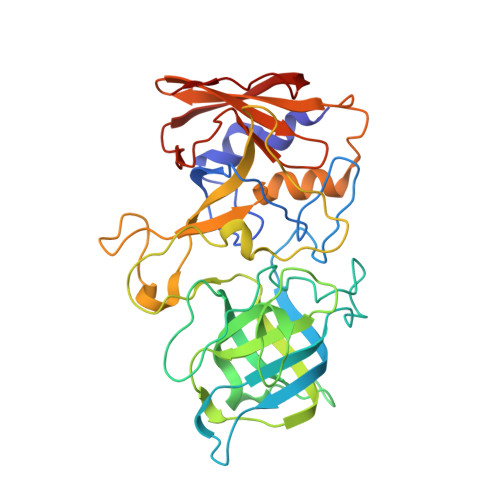
Structural analysis of a rabbit hemorrhagic disease virus binding to histo-blood group antigens.
Leuthold, M.M., Dalton, K.P., Hansman, G.S.(2015) J Virol 89: 2378-2387
- PubMed: 25505081 Search on PubMedSearch on PubMed Central
- DOI: https://doi.org/10.1128/JVI.02832-14
- Primary Citation Related Structures:
4X1W, 4X1X, 4X1Z - PubMed Abstract:
Rabbit hemorrhagic disease virus (RHDV) is a member of the Caliciviridae family (Lagovirus genus). RHDV is highly contagious and attaches to epithelial cells in the digestive or respiratory tract, leading to massive lesions with high mortality rates. A new variant of RHDV (termed RHDVb) recently has emerged, and previously vaccinated rabbits appear to have little protection against this new strain. Similar to human norovirus (Caliciviridae, Norovirus genus), RHDV binds histo-blood group antigens (HBGAs), and this is thought to be important for infection. Here, we report the HBGA binding site on the RHDVb capsid-protruding domain (P domain) using X-ray crystallography. The HBGA binding pocket was located in a negatively charged patch on the side of the P domain and at a dimeric interface. Residues from both monomers contributed to the HBGA binding and involved a network of direct hydrogen bonds and water-mediated interactions. An amino acid sequence alignment of different RHDV strains indicated that the residues directly interacting with the ABH-fucose of the HBGAs (Asp472, Asn474, and Ser479) were highly conserved. This result suggested that different RHDV strains also could bind HBGAs at the equivalent pocket. Moreover, several HBGA binding characteristics between RHDVb and human genogroup II norovirus were similar, which indicated a possible convergent evolution of HBGA binding interactions. Further structural studies with other RHDV strains are needed in order to better understand the HBGA binding mechanisms among the diverse RHDV strains. We identified, for the first time, the HBGA binding site on an RHDVb P domain using X-ray crystallography. Our results showed that RHDVb and human genogroup II noroviruses had similar HBGA binding interactions. Recently, it was discovered that synthetic HBGAs or HBGA-expressing enteric bacteria could enhance human genogroup II norovirus infection in B cells. Considering that RHDVb and genogroup II norovirus similarly interacted with HBGAs, it may be possible that a comparable cell culture system also could work with RHDVb. Taken together, these new findings will extend our understanding of calicivirus HBGA interactions and may help to elucidate the specific roles of HBGAs in calicivirus infections.
- Schaller Research Group at the University of Heidelberg and the DKFZ, Heidelberg, Germany Department of Infectious Diseases, Virology, University of Heidelberg, Heidelberg, Germany.
Organizational Affiliation: