4WZP

Ser65 phosphorylated ubiquitin, major conformation


Experimental Data Snapshot

  • Method: X-RAY DIFFRACTION
  • Resolution: 1.90 Å
  • R-Value Free: 0.237 
  • R-Value Work: 0.193 
  • R-Value Observed: 0.196 

wwPDB Validation   3D Report Full Report


This is version 2.1 of the entry. See complete history


Literature

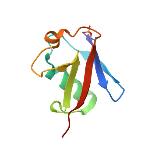
Ubiquitin Ser65 phosphorylation affects ubiquitin structure, chain assembly and hydrolysis.

Wauer, T.Swatek, K.N.Wagstaff, J.L.Gladkova, C.Pruneda, J.N.Michel, M.A.Gersch, M.Johnson, C.M.Freund, S.M.Komander, D.

(2015) EMBO J 34: 307-325

  • DOI: https://doi.org/10.15252/embj.201489847
  • Primary Citation of Related Structures:  
    4WZP

  • PubMed Abstract: 

    The protein kinase PINK1 was recently shown to phosphorylate ubiquitin (Ub) on Ser65, and phosphoUb activates the E3 ligase Parkin allosterically. Here, we show that PINK1 can phosphorylate every Ub in Ub chains. Moreover, Ser65 phosphorylation alters Ub structure, generating two conformations in solution. A crystal structure of the major conformation resembles Ub but has altered surface properties. NMR reveals a second phosphoUb conformation in which β5-strand slippage retracts the C-terminal tail by two residues into the Ub core. We further show that phosphoUb has no effect on E1-mediated E2 charging but can affect discharging of E2 enzymes to form polyUb chains. Notably, UBE2R1- (CDC34), UBE2N/UBE2V1- (UBC13/UEV1A), TRAF6- and HOIP-mediated chain assembly is inhibited by phosphoUb. While Lys63-linked poly-phosphoUb is recognized by the TAB2 NZF Ub binding domain (UBD), 10 out of 12 deubiquitinases (DUBs), including USP8, USP15 and USP30, are impaired in hydrolyzing phosphoUb chains. Hence, Ub phosphorylation has repercussions for ubiquitination and deubiquitination cascades beyond Parkin activation and may provide an independent layer of regulation in the Ub system.


  • Organizational Affiliation

    Medical Research Council Laboratory of Molecular Biology, Cambridge, UK.


Macromolecules
Find similar proteins by:  (by identity cutoff)  |  3D Structure
Entity ID: 1
MoleculeChains Sequence LengthOrganismDetailsImage
ubiquitin
A, B, C, D, E
A, B, C, D, E, F, G, H
76Homo sapiensMutation(s): 0 
Gene Names: UBB
UniProt & NIH Common Fund Data Resources
Find proteins for P0CG47 (Homo sapiens)
Explore P0CG47 
Go to UniProtKB:  P0CG47
PHAROS:  P0CG47
GTEx:  ENSG00000170315 
Entity Groups  
Sequence Clusters30% Identity50% Identity70% Identity90% Identity95% Identity100% Identity
UniProt GroupP0CG47
Sequence Annotations
Expand
  • Reference Sequence
Experimental Data & Validation

Experimental Data

  • Method: X-RAY DIFFRACTION
  • Resolution: 1.90 Å
  • R-Value Free: 0.237 
  • R-Value Work: 0.193 
  • R-Value Observed: 0.196 
  • Space Group: P 1
Unit Cell:
Length ( Å )Angle ( ˚ )
a = 49.563α = 96.86
b = 53.285β = 104.94
c = 56.866γ = 110.96
Software Package:
Software NamePurpose
PHENIXrefinement
XDSdata reduction
Aimlessdata scaling
PHASERphasing

Structure Validation

View Full Validation Report


Entry History & Funding Information

Deposition Data


Funding OrganizationLocationGrant Number
Medical Research Council (United Kingdom)United KingdomU105192732
European Research CouncilUnited Kingdom309756

Revision History  (Full details and data files)

  • Version 1.0: 2015-01-14
    Type: Initial release
  • Version 1.1: 2015-02-11
    Changes: Database references
  • Version 2.0: 2017-09-13
    Changes: Advisory, Atomic model, Author supporting evidence, Derived calculations
  • Version 2.1: 2024-01-10
    Changes: Data collection, Database references, Refinement description