4WYD

Adenosylmethionine-8-amino-7-oxononanoate aminotransferase from Mycobacterium tuberculosis complexed with a fragment from DSF screening


Experimental Data Snapshot

  • Method: X-RAY DIFFRACTION
  • Resolution: 1.35 Å
  • R-Value Free: 0.158 
  • R-Value Work: 0.126 
  • R-Value Observed: 0.127 

wwPDB Validation   3D Report Full Report


Ligand Structure Quality Assessment 


This is version 1.2 of the entry. See complete history


Literature

Fragment-Based Exploration of Binding Site Flexibility in Mycobacterium tuberculosis BioA.

Dai, R.Geders, T.W.Liu, F.Park, S.W.Schnappinger, D.Aldrich, C.C.Finzel, B.C.

(2015) J Med Chem 58: 5208-5217

  • DOI: https://doi.org/10.1021/acs.jmedchem.5b00092
  • Primary Citation of Related Structures:  
    4WYA, 4WYC, 4WYD, 4WYE, 4WYF, 4WYG, 4XEW

  • PubMed Abstract: 

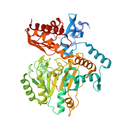
    The PLP-dependent transaminase (BioA) of Mycobacterium tuberculosis and other pathogens that catalyzes the second step of biotin biosynthesis is a now well-validated target for antibacterial development. Fragment screening by differential scanning fluorimetry has been performed to discover new chemical scaffolds and promote optimization of existing inhibitors. Calorimetry confirms binding of six molecules with high ligand efficiency. Thermodynamic data identifies which molecules bind with the enthalpy driven stabilization preferred in compounds that represent attractive starting points for future optimization. Crystallographic characterization of complexes with these molecules reveals the dynamic nature of the BioA active site. Different side chain conformational states are stabilized in response to binding by different molecules. A detailed analysis of conformational diversity in available BioA structures is presented, resulting in the identification of two states that might be targeted with molecular scaffolds incorporating well-defined conformational attributes. This new structural data can be used as part of a scaffold hopping strategy to further optimize existing inhibitors or create new small molecules with improved therapeutic potential.


  • Organizational Affiliation

    †Department of Medicinal Chemistry, University of Minnesota, 8-101 Weaver-Densford, 308 Harvard Street SE, Minneapolis, Minnesota 55455, United States.


Macromolecules
Find similar proteins by:  (by identity cutoff)  |  3D Structure
Entity ID: 1
MoleculeChains Sequence LengthOrganismDetailsImage
Adenosylmethionine-8-amino-7-oxononanoate aminotransferase
A, B
457Mycobacterium tuberculosis H37RvMutation(s): 0 
Gene Names: bioARv1568MTCY336.35c
EC: 2.6.1.62
UniProt
Find proteins for P9WQ81 (Mycobacterium tuberculosis (strain ATCC 25618 / H37Rv))
Explore P9WQ81 
Go to UniProtKB:  P9WQ81
Entity Groups  
Sequence Clusters30% Identity50% Identity70% Identity90% Identity95% Identity100% Identity
UniProt GroupP9WQ81
Sequence Annotations
Expand
  • Reference Sequence
Small Molecules
Ligands 4 Unique
IDChains Name / Formula / InChI Key2D Diagram3D Interactions
PLP
Query on PLP

Download Ideal Coordinates CCD File 
C [auth A],
M [auth B]
PYRIDOXAL-5'-PHOSPHATE
C8 H10 N O6 P
NGVDGCNFYWLIFO-UHFFFAOYSA-N
EPE
Query on EPE

Download Ideal Coordinates CCD File 
E [auth A],
O [auth B]
4-(2-HYDROXYETHYL)-1-PIPERAZINE ETHANESULFONIC ACID
C8 H18 N2 O4 S
JKMHFZQWWAIEOD-UHFFFAOYSA-N
3VR
Query on 3VR

Download Ideal Coordinates CCD File 
D [auth A],
N [auth B]
N-methyl-1-[4-(1H-pyrazol-1-ylmethyl)phenyl]methanamine
C12 H15 N3
BQLXMVISWVMAID-UHFFFAOYSA-N
EDO
Query on EDO

Download Ideal Coordinates CCD File 
F [auth A]
G [auth A]
H [auth A]
I [auth A]
J [auth A]
F [auth A],
G [auth A],
H [auth A],
I [auth A],
J [auth A],
K [auth A],
L [auth A],
P [auth B],
Q [auth B],
R [auth B],
S [auth B]
1,2-ETHANEDIOL
C2 H6 O2
LYCAIKOWRPUZTN-UHFFFAOYSA-N
Binding Affinity Annotations 
IDSourceBinding Affinity
3VR Binding MOAD:  4WYD Kd: 1.17e+4 (nM) from 1 assay(s)
Experimental Data & Validation

Experimental Data

  • Method: X-RAY DIFFRACTION
  • Resolution: 1.35 Å
  • R-Value Free: 0.158 
  • R-Value Work: 0.126 
  • R-Value Observed: 0.127 
  • Space Group: P 21 21 21
Unit Cell:
Length ( Å )Angle ( ˚ )
a = 63.104α = 90
b = 66.49β = 90
c = 204.65γ = 90
Software Package:
Software NamePurpose
PHENIXrefinement
SCALAdata scaling
PDB_EXTRACTdata extraction
XDSdata reduction
PHASERphasing

Structure Validation

View Full Validation Report


Ligand Structure Quality Assessment 


Entry History 

Deposition Data

Revision History  (Full details and data files)

  • Version 1.0: 2015-07-15
    Type: Initial release
  • Version 1.1: 2015-07-22
    Changes: Database references
  • Version 1.2: 2023-09-27
    Changes: Data collection, Database references, Derived calculations, Refinement description, Source and taxonomy