4WS2

Crystal structure of Mycobacterium tuberculosis uracil-DNA glycosylase in complex with 6-aminouracil, Form I


Experimental Data Snapshot

  • Method: X-RAY DIFFRACTION
  • Resolution: 1.13 Å
  • R-Value Free: 0.156 
  • R-Value Work: 0.128 
  • R-Value Observed: 0.129 

wwPDB Validation   3D Report Full Report


This is version 1.4 of the entry. See complete history


Literature

Structural plasticity in Mycobacterium tuberculosis uracil-DNA glycosylase (MtUng) and its functional implications.

Arif, S.M.Geethanandan, K.Mishra, P.Surolia, A.Varshney, U.Vijayan, M.

(2015) Acta Crystallogr D Biol Crystallogr 71: 1514-1527

  • DOI: https://doi.org/10.1107/S1399004715009311
  • Primary Citation of Related Structures:  
    4WPK, 4WPL, 4WRU, 4WRV, 4WRW, 4WRX, 4WRY, 4WRZ, 4WS0, 4WS1, 4WS2, 4WS3, 4WS4, 4WS5, 4WS6, 4WS7, 4WS8

  • PubMed Abstract: 

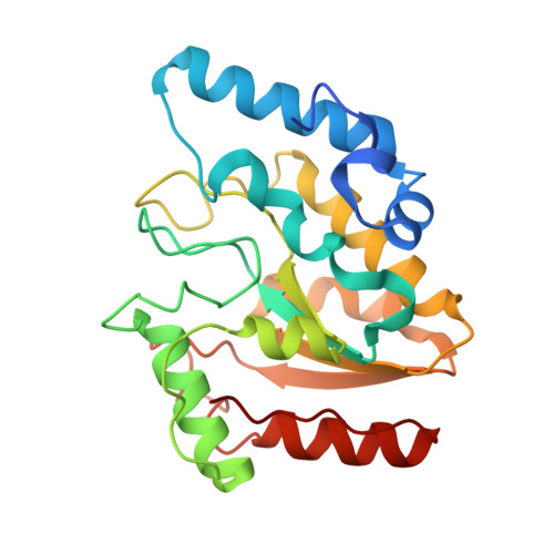
    17 independent crystal structures of family I uracil-DNA glycosylase from Mycobacterium tuberculosis (MtUng) and its complexes with uracil and its derivatives, distributed among five distinct crystal forms, have been determined. Thermodynamic parameters of binding in the complexes have been measured using isothermal titration calorimetry. The two-domain protein exhibits open and closed conformations, suggesting that the closure of the domain on DNA binding involves conformational selection. Segmental mobility in the enzyme molecule is confined to a 32-residue stretch which plays a major role in DNA binding. Uracil and its derivatives can bind to the protein in two possible orientations. Only one of them is possible when there is a bulky substituent at the 5' position. The crystal structures of the complexes provide a reasonable rationale for the observed thermodynamic parameters. In addition to providing fresh insights into the structure, plasticity and interactions of the protein molecule, the results of the present investigation provide a platform for structure-based inhibitor design.


  • Organizational Affiliation

    Molecular Biophysics Unit, Indian Institute of Science, Bangalore 560 012, India.


Macromolecules
Find similar proteins by:  (by identity cutoff)  |  3D Structure
Entity ID: 1
MoleculeChains Sequence LengthOrganismDetailsImage
Uracil-DNA glycosylase238Mycobacterium tuberculosis H37RvMutation(s): 0 
Gene Names: ungRv2976cMTCY349.11
EC: 3.2.2.27
UniProt
Find proteins for P9WFQ9 (Mycobacterium tuberculosis (strain ATCC 25618 / H37Rv))
Explore P9WFQ9 
Go to UniProtKB:  P9WFQ9
Entity Groups  
Sequence Clusters30% Identity50% Identity70% Identity90% Identity95% Identity100% Identity
UniProt GroupP9WFQ9
Sequence Annotations
Expand
  • Reference Sequence
Small Molecules
Ligands 5 Unique
IDChains Name / Formula / InChI Key2D Diagram3D Interactions
CIT
Query on CIT

Download Ideal Coordinates CCD File 
B [auth A],
C [auth A]
CITRIC ACID
C6 H8 O7
KRKNYBCHXYNGOX-UHFFFAOYSA-N
6UA
Query on 6UA

Download Ideal Coordinates CCD File 
D [auth A]6-aminopyrimidine-2,4(3H,5H)-dione
C4 H5 N3 O2
BLOCGUWRFAOSFW-UHFFFAOYSA-N
DMS
Query on DMS

Download Ideal Coordinates CCD File 
E [auth A],
F [auth A],
G [auth A],
H [auth A],
I [auth A]
DIMETHYL SULFOXIDE
C2 H6 O S
IAZDPXIOMUYVGZ-UHFFFAOYSA-N
IPA
Query on IPA

Download Ideal Coordinates CCD File 
K [auth A]ISOPROPYL ALCOHOL
C3 H8 O
KFZMGEQAYNKOFK-UHFFFAOYSA-N
CL
Query on CL

Download Ideal Coordinates CCD File 
J [auth A]CHLORIDE ION
Cl
VEXZGXHMUGYJMC-UHFFFAOYSA-M
Experimental Data & Validation

Experimental Data

  • Method: X-RAY DIFFRACTION
  • Resolution: 1.13 Å
  • R-Value Free: 0.156 
  • R-Value Work: 0.128 
  • R-Value Observed: 0.129 
  • Space Group: P 1 21 1
Unit Cell:
Length ( Å )Angle ( ˚ )
a = 38.96α = 90
b = 64.1β = 112.55
c = 45.25γ = 90
Software Package:
Software NamePurpose
REFMACrefinement
iMOSFLMdata reduction
SCALAdata scaling
PHASERphasing

Structure Validation

View Full Validation Report


Entry History 

Deposition Data

Revision History  (Full details and data files)

  • Version 1.0: 2015-07-15
    Type: Initial release
  • Version 1.1: 2015-07-22
    Changes: Database references
  • Version 1.2: 2016-04-13
    Changes: Data collection
  • Version 1.3: 2017-11-22
    Changes: Derived calculations, Refinement description
  • Version 1.4: 2023-09-27
    Changes: Data collection, Database references, Refinement description