4WJ5 | pdb_00004wj5

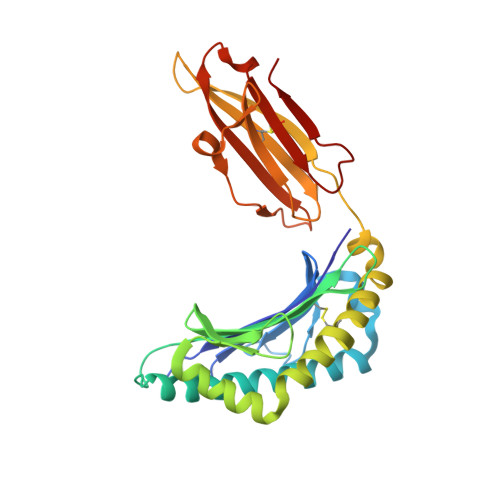
Structure of HLA-A2 in complex with an altered peptide ligands based on Mart-1 variant epitope


Experimental Data Snapshot

  • Method: X-RAY DIFFRACTION
  • Resolution: 1.65 Å
  • R-Value Free: 
    0.179 (Depositor), 0.203 (DCC) 
  • R-Value Work: 
    0.155 (Depositor), 0.176 (DCC) 
  • R-Value Observed: 
    0.156 (Depositor) 

Starting Model: experimental
View more details

wwPDB Validation 3D Report Full Report

Validation slider image for 4WJ5

This is version 1.3 of the entry. See complete history. 

Literature

Altered Peptide Ligands Revisited: Vaccine Design through Chemically Modified HLA-A2-Restricted T Cell Epitopes.

Hoppes, R., Oostvogels, R., Luimstra, J.J., Wals, K., Toebes, M., Bies, L., Ekkebus, R., Rijal, P., Celie, P.H., Huang, J.H., Emmelot, M.E., Spaapen, R.M., Lokhorst, H., Schumacher, T.N., Mutis, T., Rodenko, B., Ovaa, H.

(2014) J Immunol 193: 4803-4813

  • DOI: https://doi.org/10.4049/jimmunol.1400800
  • Primary Citation Related Structures: 
    4WJ5

  • PubMed Abstract: 

    Virus or tumor Ag-derived peptides that are displayed by MHC class I molecules are attractive starting points for vaccine development because they induce strong protective and therapeutic cytotoxic T cell responses. In thus study, we show that the MHC binding and consequent T cell reactivity against several HLA-A*02 restricted epitopes can be further improved through the incorporation of nonproteogenic amino acids at primary and secondary anchor positions. We screened more than 90 nonproteogenic, synthetic amino acids through a range of epitopes and tested more than 3000 chemically enhanced altered peptide ligands (CPLs) for binding affinity to HLA-A*0201. With this approach, we designed CPLs of viral epitopes, of melanoma-associated Ags, and of the minor histocompatibility Ag UTA2-1, which is currently being evaluated for its antileukemic activity in clinical dendritic cell vaccination trials. The crystal structure of one of the CPLs in complex with HLA-A*0201 revealed the molecular interactions likely responsible for improved binding. The best CPLs displayed enhanced affinity for MHC, increasing MHC stability and prolonging recognition by Ag-specific T cells and, most importantly, they induced accelerated expansion of antitumor T cell frequencies in vitro and in vivo as compared with the native epitope. Eventually, we were able to construct a toolbox of preferred nonproteogenic residues with which practically any given HLA-A*02 restricted epitope can be readily optimized. These CPLs could improve the therapeutic outcome of vaccination strategies or can be used for ex vivo enrichment and faster expansion of Ag-specific T cells for transfer into patients.


  • Organizational Affiliation: 
    • Division of Cell Biology, The Netherlands Cancer Institute, 1066 CX Amsterdam, the Netherlands;

Macromolecule Content 

  • Total Structure Weight: 90.63 kDa 
  • Atom Count: 7,465 
  • Modeled Residue Count: 770 
  • Deposited Residue Count: 770 
  • Unique protein chains: 3

Macromolecules

Find similar proteins by:|  3D Structure
Entity ID: 1
MoleculeChains  Sequence LengthOrganismDetailsImage
HLA class I histocompatibility antigen, A-2 alpha chain
A, D
275Homo sapiensMutation(s): 0 
Gene Names: HLA-A, HLAA
UniProt & NIH Common Fund Data Resources
Find proteins for P04439 (Homo sapiens)
Explore P04439 
Go to UniProtKB:  P04439
PHAROS:  P04439
GTEx:  ENSG00000206503 
Entity Groups
Sequence Clusters30% Identity50% Identity70% Identity90% Identity95% Identity100% Identity
UniProt GroupP04439
Sequence Annotations
Expand
Reference Sequence
Find similar proteins by:|  3D Structure
Entity ID: 2
MoleculeChains  Sequence LengthOrganismDetailsImage
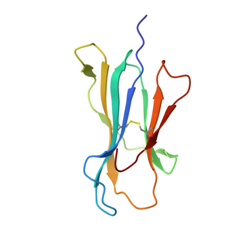
Beta-2-microglobulin
B, E
100Homo sapiensMutation(s): 0 
Gene Names: B2M, CDABP0092, HDCMA22P
UniProt & NIH Common Fund Data Resources
Find proteins for P61769 (Homo sapiens)
Explore P61769 
Go to UniProtKB:  P61769
PHAROS:  P61769
GTEx:  ENSG00000166710 
Entity Groups
Sequence Clusters30% Identity50% Identity70% Identity90% Identity95% Identity100% Identity
UniProt GroupP61769
Sequence Annotations
Expand
Reference Sequence
Find similar proteins by:  Sequence   |   3D Structure  
Entity ID: 3
MoleculeChains  Sequence LengthOrganismDetailsImage
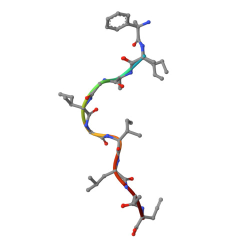
Melanoma antigen recognized by T-cells 1
C, F
10Homo sapiensMutation(s): 0 
UniProt & NIH Common Fund Data Resources
Find proteins for Q16655 (Homo sapiens)
Explore Q16655 
Go to UniProtKB:  Q16655
PHAROS:  Q16655
GTEx:  ENSG00000120215 
Entity Groups
Sequence Clusters30% Identity50% Identity70% Identity90% Identity95% Identity100% Identity
UniProt GroupQ16655
Sequence Annotations
Expand
Reference Sequence

Small Molecules

Ligands 1 Unique
IDChains Name / Formula / InChI Key2D Diagram3D Interactions
GOL

Query on GOL


Download:Ideal Coordinates CCD File
G [auth A]
H [auth A]
I [auth B]
J [auth B]
K [auth D]
G [auth A],
H [auth A],
I [auth B],
J [auth B],
K [auth D],
L [auth D],
M [auth D],
N [auth D],
O [auth D],
P [auth D],
Q [auth E],
R [auth E],
S [auth E]
GLYCEROL
C3 H8 O3
PEDCQBHIVMGVHV-UHFFFAOYSA-N
Modified Residues  2 Unique
IDChains TypeFormula2D DiagramParent
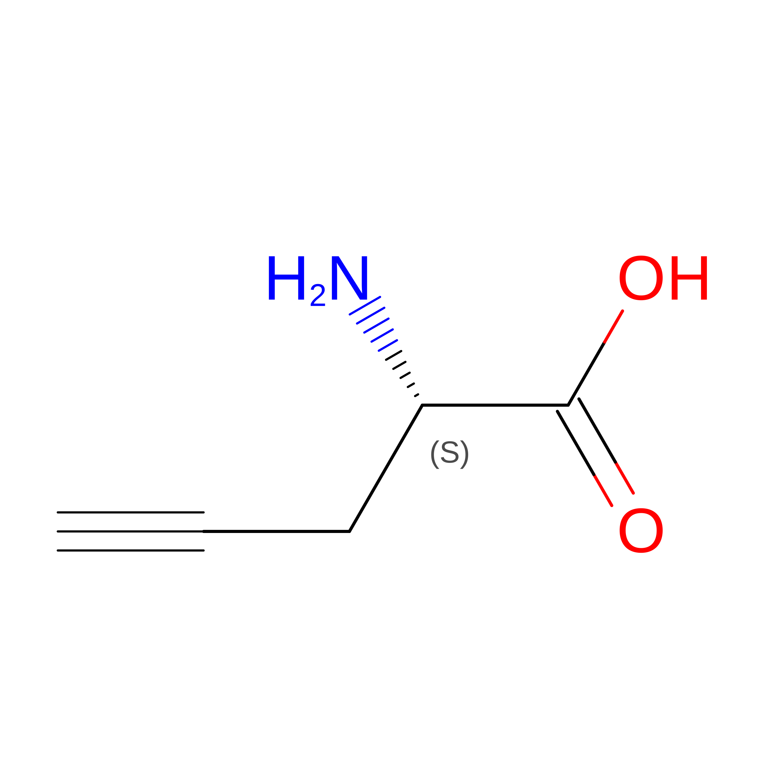
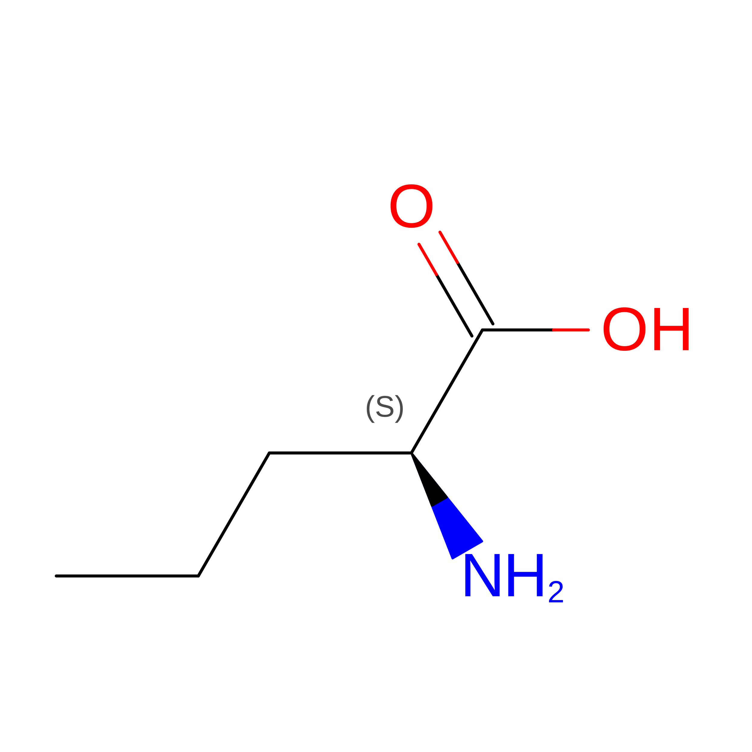
LPH
Query on LPH
C, F
L-PEPTIDE LINKINGC5 H7 N O2

--

NVA
Query on NVA
C, F
L-PEPTIDE LINKINGC5 H11 N O2VAL

Experimental Data & Validation

Experimental Data

  • Method: X-RAY DIFFRACTION
  • Resolution: 1.65 Å
  • R-Value Free:  0.179 (Depositor), 0.203 (DCC) 
  • R-Value Work:  0.155 (Depositor), 0.176 (DCC) 
  • R-Value Observed: 0.156 (Depositor) 
Space Group: P 1 21 1
Unit Cell:
Length ( Å )Angle ( ˚ )
a = 63.18α = 90
b = 87.14β = 90.15
c = 79.194γ = 90
Software Package:
Software NamePurpose
REFMACrefinement
SCALAdata scaling
XDSdata reduction
PDB_EXTRACTdata extraction
AMoREphasing

Structure Validation

View Full Validation Report


Entry History 

Deposition Data

Revision History  (Full details and data files)

  • Version 1.0: 2014-10-29
    Type: Initial release
  • Version 1.1: 2014-11-19
    Changes: Database references
  • Version 1.2: 2024-01-10
    Changes: Data collection, Database references, Derived calculations, Refinement description
  • Version 1.3: 2024-10-09
    Changes: Structure summary