4WIA | pdb_00004wia

Crystal structure of flagellar accessory protein FlaH from Methanocaldococcus jannaschii


Experimental Data Snapshot

  • Method: X-RAY DIFFRACTION
  • Resolution: 2.20 Å
  • R-Value Free: 
    0.233 (Depositor), 0.243 (DCC) 
  • R-Value Work: 
    0.206 (Depositor), 0.216 (DCC) 
  • R-Value Observed: 
    0.207 (Depositor) 

wwPDB Validation 3D Report Full Report

Validation slider image for 4WIA

This is version 1.3 of the entry. See complete history

Literature

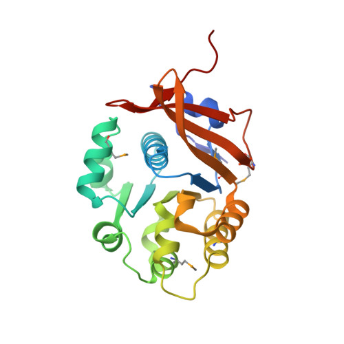
Crystal structure of the flagellar accessory protein FlaH of Methanocaldococcus jannaschii suggests a regulatory role in archaeal flagellum assembly.

Meshcheryakov, V.A.Wolf, M.

(2016) Protein Sci 25: 1147-1155

  • DOI: https://doi.org/10.1002/pro.2932
  • Primary Citation Related Structures: 
    4WIA

  • PubMed Abstract: 

    Archaeal flagella are unique structures that share functional similarity with bacterial flagella, but are structurally related to bacterial type IV pili. The flagellar accessory protein FlaH is one of the conserved components of the archaeal motility system. However, its function is not clearly understood. Here, we present the 2.2 Å resolution crystal structure of FlaH from the hyperthermophilic archaeon, Methanocaldococcus jannaschii. The protein has a characteristic RecA-like fold, which has been found previously both in archaea and bacteria. We show that FlaH binds to immobilized ATP-however, it lacks ATPase activity. Surface plasmon resonance analysis demonstrates that ATP affects the interaction between FlaH and the archaeal motor protein FlaI. In the presence of ATP, the FlaH-FlaI interaction becomes significantly weaker. A database search revealed similarity between FlaH and several DNA-binding proteins of the RecA superfamily. The closest structural homologs of FlaH are KaiC-like proteins, which are archaeal homologs of the circadian clock protein KaiC from cyanobacteria. We propose that one of the functions of FlaH may be the regulation of archaeal motor complex assembly.


  • Organizational Affiliation
    • Molecular Cryo-Electron Microscopy Unit, Okinawa Institute of Science and Technology Graduate University, 1919-1 Tancha, Onna, Kunigami, Okinawa 904-0495, Japan.

Macromolecule Content 

  • Total Structure Weight: 80.45 kDa 
  • Atom Count: 5,519 
  • Modeled Residue Count: 684 
  • Deposited Residue Count: 699 
  • Unique protein chains: 1

Macromolecules

Find similar proteins by:|  3D Structure
Entity ID: 1
MoleculeChains  Sequence LengthOrganismDetailsImage
Putative flagella-related protein H
A, B, C
233Methanocaldococcus jannaschii DSM 2661Mutation(s): 0 
Gene Names: flaHMJ0899
UniProt
Find proteins for Q58309 (Methanocaldococcus jannaschii (strain ATCC 43067 / DSM 2661 / JAL-1 / JCM 10045 / NBRC 100440))
Explore Q58309 
Go to UniProtKB:  Q58309
Entity Groups
Sequence Clusters30% Identity50% Identity70% Identity90% Identity95% Identity100% Identity
UniProt GroupQ58309
Sequence Annotations
Expand
Reference Sequence

Small Molecules

Ligands 2 Unique
IDChains Name / Formula / InChI Key2D Diagram3D Interactions
SO4

Query on SO4


Download:Ideal Coordinates CCD File
D [auth A]
E [auth A]
F [auth A]
G [auth A]
H [auth A]
D [auth A],
E [auth A],
F [auth A],
G [auth A],
H [auth A],
J [auth B],
K [auth B],
L [auth B],
M [auth B],
N [auth C],
O [auth C],
P [auth C],
Q [auth C]
SULFATE ION
O4 S
QAOWNCQODCNURD-UHFFFAOYSA-L
GOL

Query on GOL


Download:Ideal Coordinates CCD File
I [auth A]GLYCEROL
C3 H8 O3
PEDCQBHIVMGVHV-UHFFFAOYSA-N
Modified Residues  1 Unique
IDChains TypeFormula2D DiagramParent
MSE
Query on MSE
A, B, C
L-PEPTIDE LINKINGC5 H11 N O2 SeMET

Experimental Data & Validation

Experimental Data

  • Method: X-RAY DIFFRACTION
  • Resolution: 2.20 Å
  • R-Value Free:  0.233 (Depositor), 0.243 (DCC) 
  • R-Value Work:  0.206 (Depositor), 0.216 (DCC) 
  • R-Value Observed: 0.207 (Depositor) 
Space Group: P 31 2 1
Unit Cell:
Length ( Å )Angle ( ˚ )
a = 130.96α = 90
b = 130.96β = 90
c = 89.081γ = 120
Software Package:
Software NamePurpose
REFMACrefinement

Structure Validation

View Full Validation Report


Entry History 

Deposition Data

Revision History  (Full details and data files)

  • Version 1.0: 2015-10-07
    Type: Initial release
  • Version 1.1: 2016-07-20
    Changes: Database references
  • Version 1.2: 2020-02-05
    Changes: Data collection, Database references, Derived calculations
  • Version 1.3: 2024-10-30
    Changes: Data collection, Database references, Refinement description, Structure summary