4WD1 | pdb_00004wd1

Acetoacetyl-CoA Synthetase from Streptomyces lividans


Experimental Data Snapshot

  • Method: X-RAY DIFFRACTION
  • Resolution: 1.90 Å
  • R-Value Free: 
    0.194 (Depositor), 0.192 (DCC) 
  • R-Value Work: 
    0.161 (Depositor), 0.159 (DCC) 
  • R-Value Observed: 
    0.162 (Depositor) 

Starting Model: experimental
View more details

wwPDB Validation 3D Report Full Report

Validation slider image for 4WD1

This is version 1.4 of the entry. See complete history

Literature

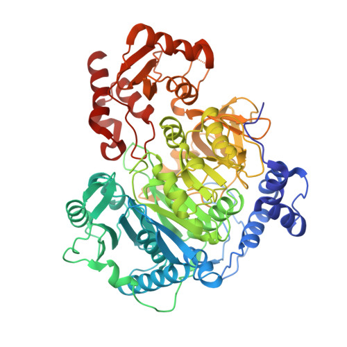
The structure of S. lividans acetoacetyl-CoA synthetase shows a novel interaction between the C-terminal extension and the N-terminal domain.

Mitchell, C.A.Tucker, A.C.Escalante-Semerena, J.C.Gulick, A.M.

(2015) Proteins 83: 575-581

  • DOI: https://doi.org/10.1002/prot.24738
  • Primary Citation Related Structures: 
    4WD1

  • PubMed Abstract: 

    The adenosine monoposphate-forming acyl-CoA synthetase enzymes catalyze a two-step reaction that involves the initial formation of an acyl adenylate that reacts in a second partial reaction to form a thioester between the acyl substrate and CoA. These enzymes utilize a Domain Alternation catalytic mechanism, whereby a ∼ 110 residue C-terminal domain rotates by 140° to form distinct catalytic conformations for the two partial reactions. The structure of an acetoacetyl-CoA synthetase (AacS) is presented that illustrates a novel aspect of this C-terminal domain. Specifically, several acetyl- and acetoacetyl-CoA synthetases contain a 30-residue extension on the C-terminus compared to other members of this family. Whereas residues from this extension are disordered in prior structures, the AacS structure shows that residues from this extension may interact with key catalytic residues from the N-terminal domain.


  • Organizational Affiliation
    • Hauptman-Woodward Institute, Buffalo, New York, 14203; Department of Structural Biology, University at Buffalo, Buffalo, New York, 14203.

Macromolecule Content 

  • Total Structure Weight: 73.78 kDa 
  • Atom Count: 5,598 
  • Modeled Residue Count: 646 
  • Deposited Residue Count: 658 
  • Unique protein chains: 1

Macromolecules

Find similar proteins by:|  3D Structure
Entity ID: 1
MoleculeChains  Sequence LengthOrganismDetailsImage
Acetoacetate-CoA ligase658Streptomyces lividans TK24Mutation(s): 0 
Gene Names: SSPG_06161

Small Molecules

Ligands 2 Unique
IDChains Name / Formula / InChI Key2D Diagram3D Interactions
EDO

Query on EDO


Download:Ideal Coordinates CCD File
B [auth A]
C [auth A]
D [auth A]
E [auth A]
F [auth A]
B [auth A],
C [auth A],
D [auth A],
E [auth A],
F [auth A],
G [auth A],
H [auth A],
I [auth A],
J [auth A],
K [auth A],
L [auth A],
M [auth A],
N [auth A],
O [auth A],
P [auth A],
Q [auth A],
R [auth A],
S [auth A],
T [auth A],
U [auth A],
V [auth A],
W [auth A]
1,2-ETHANEDIOL
C2 H6 O2
LYCAIKOWRPUZTN-UHFFFAOYSA-N
CA

Query on CA


Download:Ideal Coordinates CCD File
X [auth A],
Y [auth A]
CALCIUM ION
Ca
BHPQYMZQTOCNFJ-UHFFFAOYSA-N

Experimental Data & Validation

Experimental Data

  • Method: X-RAY DIFFRACTION
  • Resolution: 1.90 Å
  • R-Value Free:  0.194 (Depositor), 0.192 (DCC) 
  • R-Value Work:  0.161 (Depositor), 0.159 (DCC) 
  • R-Value Observed: 0.162 (Depositor) 
Space Group: P 31 2 1
Unit Cell:
Length ( Å )Angle ( ˚ )
a = 104.408α = 90
b = 104.408β = 90
c = 133.906γ = 120
Software Package:
Software NamePurpose
PHENIXrefinement
HKL-2000data scaling
PHENIXphasing

Structure Validation

View Full Validation Report


Entry History 

& Funding Information

Deposition Data


Funding OrganizationLocationGrant Number
National Institutes of Health/National Institute of General Medical Sciences (NIH/NIGMS)United StatesGM068440

Revision History  (Full details and data files)

  • Version 1.0: 2015-01-28
    Type: Initial release
  • Version 1.1: 2015-02-25
    Changes: Database references
  • Version 1.2: 2017-09-20
    Changes: Author supporting evidence, Database references, Derived calculations, Other, Refinement description, Source and taxonomy, Structure summary
  • Version 1.3: 2019-12-25
    Changes: Author supporting evidence
  • Version 1.4: 2023-09-27
    Changes: Data collection, Database references, Derived calculations, Refinement description