4WBD | pdb_00004wbd

The crystal structure of BshC from Bacillus subtilis complexed with citrate and ADP


Experimental Data Snapshot

  • Method: X-RAY DIFFRACTION
  • Resolution: 1.77 Å
  • R-Value Free: 
    0.198 (Depositor), 0.210 (DCC) 
  • R-Value Work: 
    0.168 (Depositor), 0.180 (DCC) 
  • R-Value Observed: 
    0.170 (Depositor) 

wwPDB Validation   3D Report Full Report


Ligand Structure Quality Assessment 

Created with Raphaël 2.3.0Worse 01 BetterLigand structure goodness of fit to experimental dataBest fitted ADPClick on this verticalbar to view details

This is version 1.4 of the entry. See complete history


Literature

X-ray Crystallographic Structure of BshC, a Unique Enzyme Involved in Bacillithiol Biosynthesis.

VanDuinen, A.J.Winchell, K.R.Keithly, M.E.Cook, P.D.

(2015) Biochemistry 54: 100-103

  • DOI: https://doi.org/10.1021/bi501394q
  • Primary Citation of Related Structures:  
    4WBD

  • PubMed Abstract: 

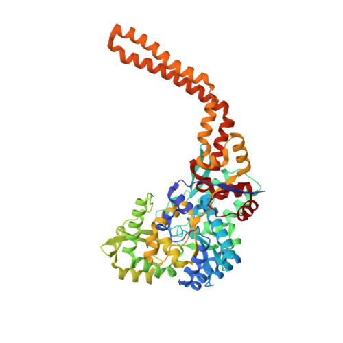
    Bacillithiol is produced by many Gram-positive bacteria via a pathway utilizing the enzymes BshA, BshB, and BshC. Here we report the 1.77 Å resolution crystal structure of BshC, the putative cysteine ligase in bacillithiol production. The structure reveals that BshC contains a core Rossmann fold with connecting peptide motifs (CP1 and CP2) and a unique α-helical coiled-coil domain that facilitates dimerization. The model contains citrate and glycerol in the canonical active site and ADP in a second binding pocket. The overall structure and bound ligands give insight into the function of this unique enzyme.


  • Organizational Affiliation

    Department of Cell & Molecular Biology, Grand Valley State University , Allendale, Michigan 49401, United States.


Macromolecules
Find similar proteins by:  (by identity cutoff)  |  3D Structure
Entity ID: 1
MoleculeChains Sequence LengthOrganismDetailsImage
BSHC541Bacillus subtilis subsp. subtilis str. 168Mutation(s): 0 
Gene Names: yllABSU15120
EC: 6
UniProt
Find proteins for P55342 (Bacillus subtilis (strain 168))
Explore P55342 
Go to UniProtKB:  P55342
Entity Groups  
Sequence Clusters30% Identity50% Identity70% Identity90% Identity95% Identity100% Identity
UniProt GroupP55342
Sequence Annotations
Expand
  • Reference Sequence
Experimental Data & Validation

Experimental Data

  • Method: X-RAY DIFFRACTION
  • Resolution: 1.77 Å
  • R-Value Free:  0.198 (Depositor), 0.210 (DCC) 
  • R-Value Work:  0.168 (Depositor), 0.180 (DCC) 
  • R-Value Observed: 0.170 (Depositor) 
Space Group: I 1 2 1
Unit Cell:
Length ( Å )Angle ( ˚ )
a = 82.53α = 90
b = 92.16β = 116.83
c = 87.377γ = 90
Software Package:
Software NamePurpose
REFMACrefinement
PDB_EXTRACTdata extraction

Structure Validation

View Full Validation Report


Ligand Structure Quality Assessment 

Created with Raphaël 2.3.0Worse 01 BetterLigand structure goodness of fit to experimental dataBest fitted ADPClick on this verticalbar to view details

Entry History & Funding Information

Deposition Data


Funding OrganizationLocationGrant Number
National Institutes of Health/National Institute of General Medical Sciences (NIH/NIGMS)United StatesF32GM093507
National Institutes of Health/National Institute of General Medical Sciences (NIH/NIGMS)United StatesR01GM030910

Revision History  (Full details and data files)

  • Version 1.0: 2014-12-31
    Type: Initial release
  • Version 1.1: 2015-01-28
    Changes: Database references
  • Version 1.2: 2017-09-13
    Changes: Author supporting evidence, Database references, Derived calculations, Other, Source and taxonomy
  • Version 1.3: 2019-12-25
    Changes: Author supporting evidence
  • Version 1.4: 2023-12-27
    Changes: Data collection, Database references, Refinement description