4V5I

Structure of the Phage P2 Baseplate in its Activated Conformation with Ca


Experimental Data Snapshot

  • Method: X-RAY DIFFRACTION
  • Resolution: 5.46 Å
  • R-Value Free: 0.297 
  • R-Value Work: 0.291 
  • R-Value Observed: 0.291 

wwPDB Validation   3D Report Full Report


This is version 1.4 of the entry. See complete history


Literature

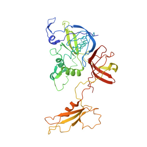
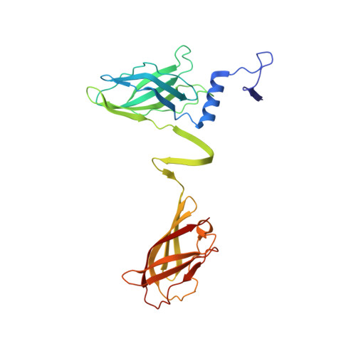
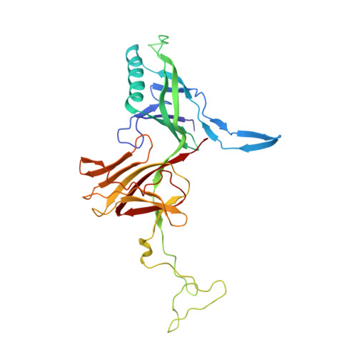
Structure of Lactococcal Phage P2 Baseplate and its Mechanism of Activation.

Sciara, G.Bebeacua, C.Bron, P.Tremblay, D.Ortiz-Lombardia, M.Lichiere, J.van Heel, M.Campanacci, V.Moineau, S.Cambillau, C.

(2010) Proc Natl Acad Sci U S A 107: 6852

  • DOI: https://doi.org/10.1073/pnas.1000232107
  • Primary Citation of Related Structures:  
    2WZP, 2X53, 4V5I

  • PubMed Abstract: 

    Siphoviridae is the most abundant viral family on earth which infects bacteria as well as archaea. All known siphophages infecting gram+ Lactococcus lactis possess a baseplate at the tip of their tail involved in host recognition and attachment. Here, we report analysis of the p2 phage baseplate structure by X-ray crystallography and electron microscopy and propose a mechanism for the baseplate activation during attachment to the host cell. This approximately 1 MDa, Escherichia coli-expressed baseplate is composed of three protein species, including six trimers of the receptor-binding protein (RBP). RBPs host-recognition domains point upwards, towards the capsid, in agreement with the electron-microscopy map of the free virion. In the presence of Ca(2+), a cation mandatory for infection, the RBPs rotated 200 degrees downwards, presenting their binding sites to the host, and a channel opens at the bottom of the baseplate for DNA passage. These conformational changes reveal a novel siphophage activation and host-recognition mechanism leading ultimately to DNA ejection.


  • Organizational Affiliation

    Architecture et Fonction des Macromolécules Biologiques, UMR 6098 Centre National de la Recherche Scientifique and Universités d'Aix-Marseille I & II, Campus de Luminy, Case 932, 13288 Marseille Cedex 09, France.


Macromolecules
Find similar proteins by:  (by identity cutoff)  |  3D Structure
Entity ID: 1
MoleculeChains Sequence LengthOrganismDetailsImage
ORF16372Lactococcus virus P2Mutation(s): 0 
Entity Groups  
Sequence Clusters30% Identity50% Identity70% Identity90% Identity95% Identity100% Identity
Sequence Annotations
Expand
  • Reference Sequence
Find similar proteins by:  (by identity cutoff)  |  3D Structure
Entity ID: 2
MoleculeChains Sequence LengthOrganismDetailsImage
PUTATIVE RECEPTOR BINDING PROTEIN263Lactococcus virus P2Mutation(s): 0 
UniProt
Find proteins for Q71AW2 (Lactococcus phage p2)
Explore Q71AW2 
Go to UniProtKB:  Q71AW2
Entity Groups  
Sequence Clusters30% Identity50% Identity70% Identity90% Identity95% Identity100% Identity
UniProt GroupQ71AW2
Sequence Annotations
Expand
  • Reference Sequence
Find similar proteins by:  (by identity cutoff)  |  3D Structure
Entity ID: 3
MoleculeChains Sequence LengthOrganismDetailsImage
ORF15298Lactococcus virus P2Mutation(s): 0 
Entity Groups  
Sequence Clusters30% Identity50% Identity70% Identity90% Identity95% Identity100% Identity
Sequence Annotations
Expand
  • Reference Sequence
Small Molecules
Ligands 1 Unique
IDChains Name / Formula / InChI Key2D Diagram3D Interactions
CA
Query on CA

Download Ideal Coordinates CCD File 
CB [auth AS]
DB [auth AT]
EB [auth AU]
FB [auth AV]
GB [auth AW]
CB [auth AS],
DB [auth AT],
EB [auth AU],
FB [auth AV],
GB [auth AW],
HB [auth AX],
IB [auth BS],
JB [auth BT],
KB [auth BU],
LB [auth BV],
MB [auth BW],
NB [auth BX]
CALCIUM ION
Ca
BHPQYMZQTOCNFJ-UHFFFAOYSA-N
Experimental Data & Validation

Experimental Data

  • Method: X-RAY DIFFRACTION
  • Resolution: 5.46 Å
  • R-Value Free: 0.297 
  • R-Value Work: 0.291 
  • R-Value Observed: 0.291 
  • Space Group: P 1 21 1
Unit Cell:
Length ( Å )Angle ( ˚ )
a = 219.521α = 90
b = 219.337β = 90.02
c = 392.432γ = 90
Software Package:
Software NamePurpose
PHASERmodel building
PHENIXrefinement
XDSdata reduction
SCALAdata scaling
PHASERphasing

Structure Validation

View Full Validation Report


Entry History 

Deposition Data

Revision History  (Full details and data files)

  • Version 1.0: 2014-07-09
    Type: Initial release
  • Version 1.1: 2014-12-10
    Changes: Other
  • Version 1.2: 2017-12-20
    Changes: Database references, Structure summary
  • Version 1.3: 2018-06-20
    Changes: Advisory, Data collection, Derived calculations
  • Version 1.4: 2024-01-10
    Changes: Data collection, Database references, Derived calculations, Refinement description