4UZK

STRUCTURE OF THE WNT DEACYLASE NOTUM FROM DROSOPHILA - CRYSTAL FORM II - 1.9A


Experimental Data Snapshot

  • Method: X-RAY DIFFRACTION
  • Resolution: 1.90 Å
  • R-Value Free: 0.204 
  • R-Value Work: 0.176 
  • R-Value Observed: 0.177 

wwPDB Validation   3D Report Full Report


This is version 1.2 of the entry. See complete history


Literature

Notum Deacylates Wnt Proteins to Suppress Signalling Activity.

Kakugawa, S.Langton, P.F.Zebisch, M.Howell, S.A.Chang, T.Liu, Y.Feizi, T.Bineva, G.O'Reilly, N.Snijders, A.P.Jones, E.Y.Vincent, J.

(2015) Nature 519: 187

  • DOI: https://doi.org/10.1038/nature14259
  • Primary Citation of Related Structures:  
    4UYU, 4UYW, 4UYZ, 4UZ1, 4UZ5, 4UZ6, 4UZ7, 4UZ9, 4UZA, 4UZJ, 4UZK, 4UZL, 4UZQ, 4WBH

  • PubMed Abstract: 

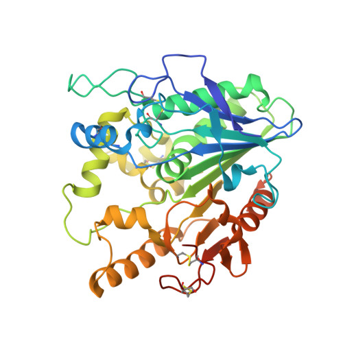
    Signalling by Wnt proteins is finely balanced to ensure normal development and tissue homeostasis while avoiding diseases such as cancer. This is achieved in part by Notum, a highly conserved secreted feedback antagonist. Notum has been thought to act as a phospholipase, shedding glypicans and associated Wnt proteins from the cell surface. However, this view fails to explain specificity, as glypicans bind many extracellular ligands. Here we provide genetic evidence in Drosophila that Notum requires glypicans to suppress Wnt signalling, but does not cleave their glycophosphatidylinositol anchor. Structural analyses reveal glycosaminoglycan binding sites on Notum, which probably help Notum to co-localize with Wnt proteins. They also identify, at the active site of human and Drosophila Notum, a large hydrophobic pocket that accommodates palmitoleate. Kinetic and mass spectrometric analyses of human proteins show that Notum is a carboxylesterase that removes an essential palmitoleate moiety from Wnt proteins and thus constitutes the first known extracellular protein deacylase.


  • Organizational Affiliation

    MRC's National Institute for Medical Research, The Ridgeway, Mill Hill, London NW7 1AA, UK.


Macromolecules
Find similar proteins by:  (by identity cutoff)  |  3D Structure
Entity ID: 1
MoleculeChains Sequence LengthOrganismDetailsImage
NOTUM
A, B
373Drosophila melanogasterMutation(s): 0 
EC: 3.1.1.1
UniProt
Find proteins for Q9VUX3 (Drosophila melanogaster)
Explore Q9VUX3 
Go to UniProtKB:  Q9VUX3
Entity Groups  
Sequence Clusters30% Identity50% Identity70% Identity90% Identity95% Identity100% Identity
UniProt GroupQ9VUX3
Sequence Annotations
Expand
  • Reference Sequence
Small Molecules
Ligands 1 Unique
IDChains Name / Formula / InChI Key2D Diagram3D Interactions
NAG
Query on NAG

Download Ideal Coordinates CCD File 
C [auth A],
D [auth B]
2-acetamido-2-deoxy-beta-D-glucopyranose
C8 H15 N O6
OVRNDRQMDRJTHS-FMDGEEDCSA-N
Experimental Data & Validation

Experimental Data

  • Method: X-RAY DIFFRACTION
  • Resolution: 1.90 Å
  • R-Value Free: 0.204 
  • R-Value Work: 0.176 
  • R-Value Observed: 0.177 
  • Space Group: P 21 21 21
Unit Cell:
Length ( Å )Angle ( ˚ )
a = 59.252α = 90
b = 93.103β = 90
c = 142.124γ = 90
Software Package:
Software NamePurpose
REFMACrefinement
XDSdata reduction
Aimlessdata scaling

Structure Validation

View Full Validation Report


Entry History 

Deposition Data

Revision History  (Full details and data files)

  • Version 1.0: 2015-02-25
    Type: Initial release
  • Version 1.1: 2015-03-18
    Changes: Database references
  • Version 1.2: 2020-07-29
    Type: Remediation
    Reason: Carbohydrate remediation
    Changes: Data collection, Derived calculations, Other, Structure summary