4US5 | pdb_00004us5

Crystal Structure of apo-MsnO8


Experimental Data Snapshot

  • Method: X-RAY DIFFRACTION
  • Resolution: 1.80 Å
  • R-Value Free: 
    0.216 (Depositor), 0.228 (DCC) 
  • R-Value Work: 
    0.188 (Depositor), 0.201 (DCC) 
  • R-Value Observed: 
    0.189 (Depositor) 

wwPDB Validation 3D Report Full Report

Validation slider image for 4US5

Ligand Structure Quality Assessment 


This is version 1.2 of the entry. See complete history

Literature

Insights Into the Bioactivity of Mensacarcin and Epoxide Formation by Msno8.

Maier, S.Pflueger, T.Loesgen, S.Asmus, K.Broetz, E.Paululat, T.Zeeck, A.Andrade, S.Bechthold, A.

(2014) Chembiochem 15: 749

  • DOI: https://doi.org/10.1002/cbic.201300704
  • Primary Citation Related Structures: 
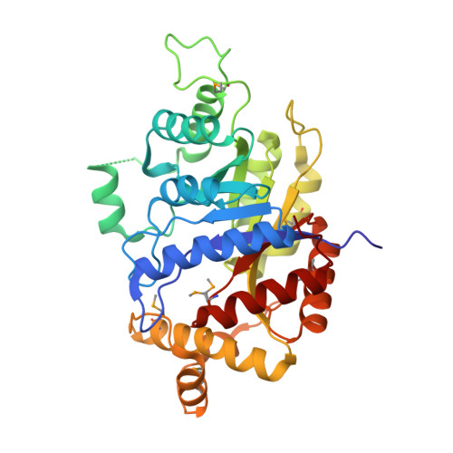
    4US5

  • PubMed Abstract: 

    Mensacarcin, a potential antitumour drug, is produced by Streptomyces bottropensis. The structure consists of a three-membered ring system with many oxygen atoms. Of vital importance in this context is an epoxy moiety in the side chain of mensacarcin. Our studies with different mensacarcin derivatives have demonstrated that this epoxy group is primarily responsible for the cytotoxic effect of mensacarcin. In order to obtain further information about this epoxy moiety, inactivation experiments in the gene cluster were carried out to identify the epoxy-forming enzyme. Therefore the cosmid cos2, which covers almost the complete type II polyketide synthase (PKS) gene cluster, was heterologously expressed in Streptomyces albus. This led to production of didesmethylmensacarcin, due to the fact that methyltransferase genes are missing in the cosmid. Further gene inactivation experiments on this cosmid showed that MsnO8, a luciferase-like monooxygenase, introduces the epoxy group at the end of the biosynthesis of mensacarcin. In addition, the protein MsnO8 was purified, and its crystal structure was determined to a resolution of 1.80 Å.


  • Organizational Affiliation
    • Institut für Pharmazeutische Biologie und Biotechnologie, Albert-Ludwigs Universität, Stefan-Meier-Strasse 19, 79104 Freiburg (Germany).

Macromolecule Content 

  • Total Structure Weight: 152.96 kDa 
  • Atom Count: 11,466 
  • Modeled Residue Count: 1,323 
  • Deposited Residue Count: 1,352 
  • Unique protein chains: 2

Macromolecules

Find similar proteins by:|  3D Structure
Entity ID: 1
MoleculeChains  Sequence LengthOrganismDetailsImage
LUCIFERASE-LIKE MONOOXYGENASE
A, B, C
338Streptomyces bottropensisMutation(s): 0 
UniProt
Find proteins for W8QCH3 (Streptomyces bottropensis)
Explore W8QCH3 
Go to UniProtKB:  W8QCH3
Entity Groups
Sequence Clusters30% Identity50% Identity70% Identity90% Identity95% Identity100% Identity
UniProt GroupW8QCH3
Sequence Annotations
Expand
Reference Sequence
Find similar proteins by:|  3D Structure
Entity ID: 2
MoleculeChains  Sequence LengthOrganismDetailsImage
LUCIFERASE-LIKE MONOOXYGENASE338Streptomyces bottropensisMutation(s): 0 
UniProt
Find proteins for W8QCH3 (Streptomyces bottropensis)
Explore W8QCH3 
Go to UniProtKB:  W8QCH3
Entity Groups
Sequence Clusters30% Identity50% Identity70% Identity90% Identity95% Identity100% Identity
UniProt GroupW8QCH3
Sequence Annotations
Expand
Reference Sequence

Small Molecules

Ligands 5 Unique
IDChains Name / Formula / InChI Key2D Diagram3D Interactions
EPE

Query on EPE


Download:Ideal Coordinates CCD File
BA [auth C],
K [auth A],
LA [auth D],
S [auth B]
4-(2-HYDROXYETHYL)-1-PIPERAZINE ETHANESULFONIC ACID
C8 H18 N2 O4 S
JKMHFZQWWAIEOD-UHFFFAOYSA-N
BGC

Query on BGC


Download:Ideal Coordinates CCD File
CA [auth C]beta-D-glucopyranose
C6 H12 O6
WQZGKKKJIJFFOK-VFUOTHLCSA-N
P4G

Query on P4G


Download:Ideal Coordinates CCD File
DA [auth C],
EA [auth C],
MA [auth D]
1-ETHOXY-2-(2-ETHOXYETHOXY)ETHANE
C8 H18 O3
RRQYJINTUHWNHW-UHFFFAOYSA-N
CL

Query on CL


Download:Ideal Coordinates CCD File
AA [auth C]CHLORIDE ION
Cl
VEXZGXHMUGYJMC-UHFFFAOYSA-M
NA

Query on NA


Download:Ideal Coordinates CCD File
E [auth A]
F [auth A]
FA [auth D]
G [auth A]
GA [auth D]
E [auth A],
F [auth A],
FA [auth D],
G [auth A],
GA [auth D],
H [auth A],
HA [auth D],
I [auth A],
IA [auth D],
J [auth A],
JA [auth D],
KA [auth D],
L [auth B],
M [auth B],
N [auth B],
O [auth B],
P [auth B],
Q [auth B],
R [auth B],
T [auth C],
U [auth C],
V [auth C],
W [auth C],
X [auth C],
Y [auth C],
Z [auth C]
SODIUM ION
Na
FKNQFGJONOIPTF-UHFFFAOYSA-N
Modified Residues  1 Unique
IDChains TypeFormula2D DiagramParent
MSE
Query on MSE
A, B, C
L-PEPTIDE LINKINGC5 H11 N O2 SeMET

Experimental Data & Validation

Experimental Data

  • Method: X-RAY DIFFRACTION
  • Resolution: 1.80 Å
  • R-Value Free:  0.216 (Depositor), 0.228 (DCC) 
  • R-Value Work:  0.188 (Depositor), 0.201 (DCC) 
  • R-Value Observed: 0.189 (Depositor) 
Space Group: P 21 21 21
Unit Cell:
Length ( Å )Angle ( ˚ )
a = 85.005α = 90
b = 102.902β = 90
c = 160.697γ = 90
Software Package:
Software NamePurpose
REFMACrefinement
XDSdata reduction
XSCALEdata scaling

Structure Validation

View Full Validation Report


Ligand Structure Quality Assessment 


Entry History 

Deposition Data

Revision History  (Full details and data files)

  • Version 1.0: 2014-07-16
    Type: Initial release
  • Version 1.1: 2020-07-29
    Type: Remediation
    Reason: Carbohydrate remediation
    Changes: Data collection, Derived calculations, Other, Structure summary
  • Version 1.2: 2024-11-20
    Changes: Data collection, Database references, Derived calculations, Structure summary