4UQI

AP2 controls clathrin polymerization with a membrane-activated switch


Experimental Data Snapshot

  • Method: X-RAY DIFFRACTION
  • Resolution: 2.79 Å
  • R-Value Free: 0.259 
  • R-Value Work: 0.203 
  • R-Value Observed: 0.206 

Starting Model: experimental
View more details

wwPDB Validation   3D Report Full Report


Ligand Structure Quality Assessment 


This is version 1.3 of the entry. See complete history


Literature

Clathrin Adaptors. Ap2 Controls Clathrin Polymerization with a Membrane-Activated Switch.

Kelly, B.T.Graham, S.C.Liska, N.Dannhauser, P.N.Honing, S.Ungewickell, E.J.Owen, D.J.

(2014) Science 345: 459

  • DOI: https://doi.org/10.1126/science.1254836
  • Primary Citation of Related Structures:  
    4UQI

  • PubMed Abstract: 

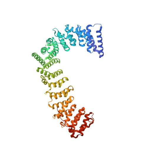
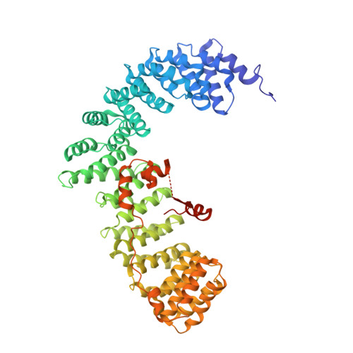
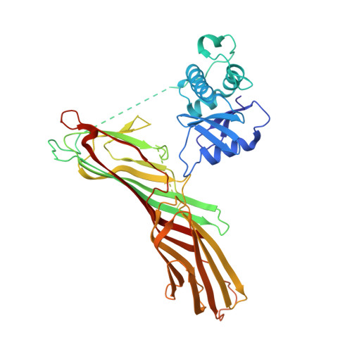
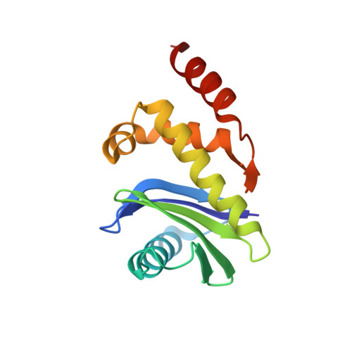
    Clathrin-mediated endocytosis (CME) is vital for the internalization of most cell-surface proteins. In CME, plasma membrane-binding clathrin adaptors recruit and polymerize clathrin to form clathrin-coated pits into which cargo is sorted. Assembly polypeptide 2 (AP2) is the most abundant adaptor and is pivotal to CME. Here, we determined a structure of AP2 that includes the clathrin-binding β2 hinge and developed an AP2-dependent budding assay. Our findings suggest that an autoinhibitory mechanism prevents clathrin recruitment by cytosolic AP2. A large-scale conformational change driven by the plasma membrane phosphoinositide phosphatidylinositol 4,5-bisphosphate and cargo relieves this autoinhibition, triggering clathrin recruitment and hence clathrin-coated bud formation. This molecular switching mechanism can couple AP2's membrane recruitment to its key functions of cargo and clathrin binding.


  • Organizational Affiliation

    Cambridge Institute for Medical Research (CIMR), Department of Clinical Biochemistry, University of Cambridge, Hills Road, Cambridge CB2 0XY, UK. btk1000@cam.ac.uk djo30@cam.ac.uk.


Macromolecules
Find similar proteins by:  (by identity cutoff)  |  3D Structure
Entity ID: 1
MoleculeChains Sequence LengthOrganismDetailsImage
AP-2 COMPLEX SUBUNIT ALPHA-2628Rattus norvegicusMutation(s): 0 
UniProt
Find proteins for P18484 (Rattus norvegicus)
Explore P18484 
Go to UniProtKB:  P18484
Entity Groups  
Sequence Clusters30% Identity50% Identity70% Identity90% Identity95% Identity100% Identity
UniProt GroupP18484
Sequence Annotations
Expand
  • Reference Sequence
Find similar proteins by:  (by identity cutoff)  |  3D Structure
Entity ID: 2
MoleculeChains Sequence LengthOrganismDetailsImage
AP-2 COMPLEX SUBUNIT BETA657Homo sapiensMutation(s): 0 
UniProt & NIH Common Fund Data Resources
Find proteins for P63010 (Homo sapiens)
Explore P63010 
Go to UniProtKB:  P63010
GTEx:  ENSG00000006125 
Entity Groups  
Sequence Clusters30% Identity50% Identity70% Identity90% Identity95% Identity100% Identity
UniProt GroupP63010
Sequence Annotations
Expand
  • Reference Sequence
Find similar proteins by:  (by identity cutoff)  |  3D Structure
Entity ID: 3
MoleculeChains Sequence LengthOrganismDetailsImage
AP-2 COMPLEX SUBUNIT MUC [auth M]446Rattus norvegicusMutation(s): 0 
UniProt
Find proteins for P84092 (Rattus norvegicus)
Explore P84092 
Go to UniProtKB:  P84092
Entity Groups  
Sequence Clusters30% Identity50% Identity70% Identity90% Identity95% Identity100% Identity
UniProt GroupP84092
Sequence Annotations
Expand
  • Reference Sequence
Find similar proteins by:  (by identity cutoff)  |  3D Structure
Entity ID: 4
MoleculeChains Sequence LengthOrganismDetailsImage
AP-2 COMPLEX SUBUNIT SIGMAD [auth S]142Mus musculusMutation(s): 0 
UniProt & NIH Common Fund Data Resources
Find proteins for P62743 (Mus musculus)
Explore P62743 
Go to UniProtKB:  P62743
IMPC:  MGI:2141861
Entity Groups  
Sequence Clusters30% Identity50% Identity70% Identity90% Identity95% Identity100% Identity
UniProt GroupP62743
Sequence Annotations
Expand
  • Reference Sequence
Small Molecules
Ligands 2 Unique
IDChains Name / Formula / InChI Key2D Diagram3D Interactions
IHP
Query on IHP

Download Ideal Coordinates CCD File 
F [auth A]INOSITOL HEXAKISPHOSPHATE
C6 H18 O24 P6
IMQLKJBTEOYOSI-GPIVLXJGSA-N
CL
Query on CL

Download Ideal Coordinates CCD File 
E [auth A]CHLORIDE ION
Cl
VEXZGXHMUGYJMC-UHFFFAOYSA-M
Experimental Data & Validation

Experimental Data

  • Method: X-RAY DIFFRACTION
  • Resolution: 2.79 Å
  • R-Value Free: 0.259 
  • R-Value Work: 0.203 
  • R-Value Observed: 0.206 
  • Space Group: P 31 2 1
Unit Cell:
Length ( Å )Angle ( ˚ )
a = 121.3α = 90
b = 121.3β = 90
c = 259.37γ = 120
Software Package:
Software NamePurpose
REFMACrefinement
XDSdata reduction
SCALAdata scaling

Structure Validation

View Full Validation Report


Ligand Structure Quality Assessment 


Entry History 

Deposition Data

Revision History  (Full details and data files)

  • Version 1.0: 2014-07-30
    Type: Initial release
  • Version 1.1: 2014-08-06
    Changes: Database references
  • Version 1.2: 2020-10-07
    Changes: Derived calculations, Other, Structure summary
  • Version 1.3: 2024-01-10
    Changes: Data collection, Database references, Refinement description