First attempts to crystallize a non-homogeneous sample of thioredoxin from Litopenaeus vannamei: What to do when you have diffraction data of a protein that is not the target?
Campos-Acevedo, A.A., Diaz-Vilchis, A., Sotelo-Mundo, R.R., Rudino-Pinera, E.(2016) Biochem Biophys Rep 8: 284-289
- PubMed: 28955968 Search on PubMedSearch on PubMed Central
- DOI: https://doi.org/10.1016/j.bbrep.2016.09.011
- Primary Citation Related Structures:
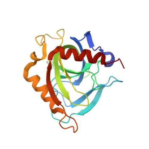
4UM4 - PubMed Abstract:
The importance of sample homogeneity and purity in protein crystallization is essential to obtain high-quality diffracting crystals. Here, in an attempt to determine the crystal structure of thioredoxin 1 from whiteleg shrimp Litopenaeus vannamei ( Lv Trx), we inadvertently crystallized the hexameric inorganic pyrophosphatase of Escherichia coli (E-PPase) from a non-homogeneous sample product during the initial over-expression steps and partial purification of Lv Trx. The structure determination and identification of the crystallized protein were derived from several clues: the failures in the Molecular Replacement (MR) trials using Lv Trx coordinates as a search model, the unit cell parameters and space group determination, and essentially by the use of the program BALBES . After using the previously deposited E-PPase structure (PDB entry 1mjw) as a search model and the correct space group assignation, the MR showed an E-PPase complexed with SO 4 -2 with small changes in the sulfate ion binding region when it compares to previously deposited E-PPases in the PDB. This work stresses the importance of protein purity to avoid the risk of crystallizing a contaminant protein or how pure need to be a protein sample in order to increase the possibility to obtain crystals, but also serves as a reminder that crystallization is by itself a purification process and how the program BALBES can be useful in the crystal structure determination of previously deposited structures in the PDB.
- Departamento de Medicina Molecular y Bioprocesos, Instituto de Biotecnología (IBT), Universidad Nacional Autónoma de México (UNAM), Avenida Universidad 2001, Colonia Chamilpa, PO Box 62210, Cuernavaca, Morelos, Mexico.
Organizational Affiliation: