4UFR | pdb_00004ufr

Structure of the ectodomain of LGR5 in complex with R-spondin-2 (Fu1Fu2)


Experimental Data Snapshot

  • Method: X-RAY DIFFRACTION
  • Resolution: 2.20 Å
  • R-Value Free: 
    0.262 (Depositor), 0.258 (DCC) 
  • R-Value Work: 
    0.208 (Depositor), 0.212 (DCC) 
  • R-Value Observed: 
    0.209 (Depositor) 

wwPDB Validation 3D Report Full Report

Validation slider image for 4UFR

Ligand Structure Quality Assessment 


This is version 1.5 of the entry. See complete history

Literature

Crystal Structure of R-Spondin 2 in Complex with the Ectodomains of its Receptors Lgr5 and Znrf3.

Zebisch, M.Jones, E.Y.

(2015) J Struct Biol 191: 149

  • DOI: https://doi.org/10.1016/j.jsb.2015.05.008
  • Primary Citation Related Structures: 
    4UFR, 4UFS

  • PubMed Abstract: 

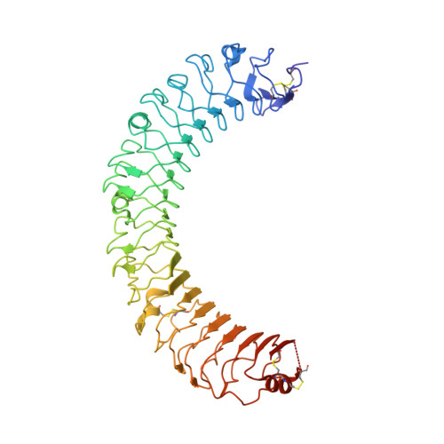
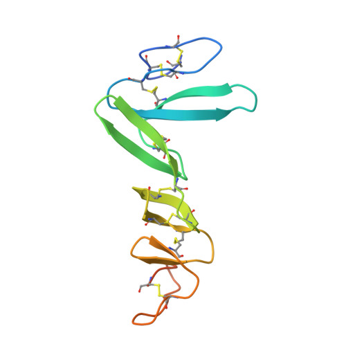
    The four secreted R-spondin (Rspo1-4) proteins of vertebrates function as stem cell growth factors and potentiate canonical Wnt signalling. Rspo proteins act by cross-linking members of two cell surface receptor families, complexing the stem cell markers LGR4-6 with the Frizzled-specific E3 ubiquitin ligases ZNRF3/RNF43. The consequent internalisation of the ternary LGR-Rspo-E3 complex removes the E3 ligase activity, which otherwise targets the Wnt receptor Frizzled for degradation, and thus enhances Wnt signalling. Multiple combinations of LGR4-6, Rspo1-4 and ZNRF3/RNF43 are possible, implying the existence of generic interaction determinants, but also of specific differences in complex architecture and activity. We present here a high resolution crystal structure of an ectodomain variant of human LGR5 (hLGR5ecto) complexed with a signalling competent fragment of mouse Rspo2 (mRspo2Fu1-Fu2). The structure shows that the particularly potent Rspo2 ligand engages LGR5 in a fashion almost identical to that reported for hRSPO1. Comparison of our hLGR5ecto structure with previously published structures highlights a surprising plasticity of the LGR ectodomains, characterised by a nearly 9° or larger rotation of the N-terminal half of the horseshoe-like fold relative to the C-terminal half. We also report a low resolution hLGR5-mRspo2Fu1-Fu2-mZNRF3ecto ternary complex structure. This crystal structure confirms our previously suggested hypothesis, showing that Rspo proteins cross-link LGRs and ZNRF3 into a 2:2:2 complex, whereas a 1:1:1 complex is formed with RNF43.


  • Organizational Affiliation
    • Division of Structural Biology, Henry Wellcome Trust Centre for Human Genetics, University of Oxford, Oxford OX3 7BN, United Kingdom.

Macromolecule Content 

  • Total Structure Weight: 136.65 kDa 
  • Atom Count: 8,912 
  • Modeled Residue Count: 1,121 
  • Deposited Residue Count: 1,208 
  • Unique protein chains: 2

Macromolecules

Find similar proteins by:|  3D Structure
Entity ID: 1
MoleculeChains  Sequence LengthOrganismDetailsImage
LEUCINE-RICH REPEAT-CONTAINING G-PROTEIN COUPLED RECEPTOR 5
A, C
484Homo sapiensMutation(s): 0 
UniProt & NIH Common Fund Data Resources
Find proteins for O75473 (Homo sapiens)
Explore O75473 
Go to UniProtKB:  O75473
PHAROS:  O75473
GTEx:  ENSG00000139292 
Entity Groups
Sequence Clusters30% Identity50% Identity70% Identity90% Identity95% Identity100% Identity
UniProt GroupO75473
Glycosylation
Glycosylation Sites: 1Go to GlyGen: O75473-1
Sequence Annotations
Expand
Reference Sequence
Find similar proteins by:|  3D Structure
Entity ID: 2
MoleculeChains  Sequence LengthOrganismDetailsImage
R-SPONDIN-2
B, D
120Homo sapiensMutation(s): 0 
UniProt
Find proteins for Q8BFU0 (Mus musculus)
Explore Q8BFU0 
Go to UniProtKB:  Q8BFU0
Entity Groups
Sequence Clusters30% Identity50% Identity70% Identity90% Identity95% Identity100% Identity
UniProt GroupQ8BFU0
Sequence Annotations
Expand
Reference Sequence

Small Molecules

Ligands 2 Unique
IDChains Name / Formula / InChI Key2D Diagram3D Interactions
NAG

Query on NAG


Download:Ideal Coordinates CCD File
E [auth A],
J [auth C]
2-acetamido-2-deoxy-beta-D-glucopyranose
C8 H15 N O6
OVRNDRQMDRJTHS-FMDGEEDCSA-N
CL

Query on CL


Download:Ideal Coordinates CCD File
F [auth A]
G [auth A]
H [auth A]
I [auth B]
K [auth C]
F [auth A],
G [auth A],
H [auth A],
I [auth B],
K [auth C],
L [auth D]
CHLORIDE ION
Cl
VEXZGXHMUGYJMC-UHFFFAOYSA-M

Experimental Data & Validation

Experimental Data

  • Method: X-RAY DIFFRACTION
  • Resolution: 2.20 Å
  • R-Value Free:  0.262 (Depositor), 0.258 (DCC) 
  • R-Value Work:  0.208 (Depositor), 0.212 (DCC) 
  • R-Value Observed: 0.209 (Depositor) 
Space Group: C 1 2 1
Unit Cell:
Length ( Å )Angle ( ˚ )
a = 205.148α = 90
b = 59.777β = 99.51
c = 112.21γ = 90
Software Package:
Software NamePurpose
REFMACrefinement

Structure Validation

View Full Validation Report


Ligand Structure Quality Assessment 


Entry History 

Deposition Data

Revision History  (Full details and data files)

  • Version 1.0: 2015-07-08
    Type: Initial release
  • Version 1.1: 2015-07-15
    Changes: Structure summary
  • Version 1.2: 2015-08-12
    Changes: Database references
  • Version 1.3: 2015-09-23
    Changes: Database references
  • Version 1.4: 2020-07-29
    Type: Remediation
    Reason: Carbohydrate remediation
    Changes: Data collection, Derived calculations, Other, Structure summary
  • Version 1.5: 2024-11-13
    Changes: Data collection, Database references, Structure summary