4UFG | pdb_00004ufg

Thrombin in complex with (2R)-2-(benzylsulfonylamino)-N-((1S)-2-((4- carbamimidoylphenyl)methylamino)-1-methyl-2-oxo-ethyl)-N-methyl-3- phenyl-propanamide ethane


Experimental Data Snapshot

  • Method: X-RAY DIFFRACTION
  • Resolution: 1.65 Å
  • R-Value Free: 
    0.186 (Depositor), 0.186 (DCC) 
  • R-Value Work: 
    0.162 (Depositor), 0.162 (DCC) 
  • R-Value Observed: 
    0.163 (Depositor) 

Starting Model: experimental
View more details

wwPDB Validation 3D Report Full Report

Validation slider image for 4UFG

Ligand Structure Quality Assessment 


This is version 1.4 of the entry. See complete history

Literature

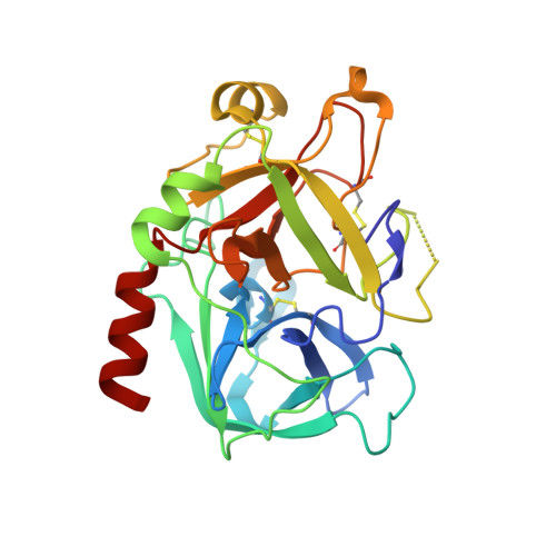
Boosting Affinity by Correct Ligand Preorganization for the S2 Pocket of Thrombin: A Study by Isothermal Titration Calorimetry, Molecular Dynamics, and High-Resolution Crystal Structures.

Ruhmann, E.H.Rupp, M.Betz, M.Heine, A.Klebe, G.

(2016) ChemMedChem 11: 309

  • DOI: https://doi.org/10.1002/cmdc.201500531
  • Primary Citation Related Structures: 
    4UFD, 4UFE, 4UFF, 4UFG

  • PubMed Abstract: 

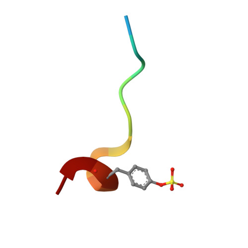
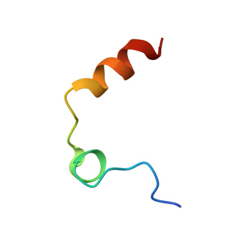
    Structural preorganization to fix bioactive conformations at protein binding sites is a popular strategy to enhance binding affinity during late-stage optimization. The rationale for this enhancement relates to entropic advantages assigned to rigidified versus flexible ligands. We analyzed a narrow series of peptidomimetics binding to thrombin. The individual ligands exhibit at P2 a conformationally flexible glycine, more restricted alanine, N-methylglycine, N-methylhomoalanine, and largely rigidified proline moiety. Overall, affinity was found to increase by a factor of 1000, explained partly by an entropic advantage. All ligands adopt the same binding mode with small deviations. The residual mobility of the bound ligands is decreased across the series, and a protein side chain differs in its order/disorder behavior along with changes in the surface-water network pattern established across the newly generated protein-ligand surfaces. The enthalpy/entropy inventory displays a rather complex picture and emphasizes that thermodynamics can only be compared in terms of relative differences within a structurally similar ligand series.


  • Organizational Affiliation
    • Institute of Pharmaceutical Chemistry, Philipps University Marburg, Marbacher Weg 6, 35037, Marburg, Germany.

Macromolecule Content 

  • Total Structure Weight: 35.54 kDa 
  • Atom Count: 2,672 
  • Modeled Residue Count: 291 
  • Deposited Residue Count: 299 
  • Unique protein chains: 3

Macromolecules

Find similar proteins by:|  3D Structure
Entity ID: 1
MoleculeChains  Sequence LengthOrganismDetailsImage
THROMBIN HEAVY CHAINA [auth H]258Homo sapiensMutation(s): 0 
EC: 3.4.21.5
UniProt & NIH Common Fund Data Resources
Find proteins for P00734 (Homo sapiens)
Explore P00734 
Go to UniProtKB:  P00734
PHAROS:  P00734
GTEx:  ENSG00000180210 
Entity Groups
Sequence Clusters30% Identity50% Identity70% Identity90% Identity95% Identity100% Identity
UniProt GroupP00734
Glycosylation
Glycosylation Sites: 1Go to GlyGen: P00734-1
Sequence Annotations
Expand
Reference Sequence
Find similar proteins by:  Sequence   |   3D Structure  
Entity ID: 2
MoleculeChains  Sequence LengthOrganismDetailsImage
HIRUDIN VARIANT-2B [auth I]12Hirudo medicinalisMutation(s): 0 
UniProt
Find proteins for P09945 (Hirudo medicinalis)
Explore P09945 
Go to UniProtKB:  P09945
Entity Groups
Sequence Clusters30% Identity50% Identity70% Identity90% Identity95% Identity100% Identity
UniProt GroupP09945
Sequence Annotations
Expand
Reference Sequence
Find similar proteins by:|  3D Structure
Entity ID: 3
MoleculeChains  Sequence LengthOrganismDetailsImage
THROMBIN LIGHT CHAINC [auth L]29Homo sapiensMutation(s): 0 
EC: 3.4.21.5
UniProt & NIH Common Fund Data Resources
Find proteins for P00734 (Homo sapiens)
Explore P00734 
Go to UniProtKB:  P00734
PHAROS:  P00734
GTEx:  ENSG00000180210 
Entity Groups
Sequence Clusters30% Identity50% Identity70% Identity90% Identity95% Identity100% Identity
UniProt GroupP00734
Sequence Annotations
Expand
Reference Sequence

Small Molecules

Ligands 4 Unique
IDChains Name / Formula / InChI Key2D Diagram3D Interactions
D6J

Query on D6J


Download:Ideal Coordinates CCD File
F [auth H](2S)-N-[(4-carbamimidoylphenyl)methyl]-2-[methyl-[(2R)-3-phenyl-2-[(phenylmethyl)sulfonylamino]propanoyl]amino]butanamide
C29 H35 N5 O4 S
OVCZZMWCFCLIPI-FTJBHMTQSA-N
NAG

Query on NAG


Download:Ideal Coordinates CCD File
H
2-acetamido-2-deoxy-beta-D-glucopyranose
C8 H15 N O6
OVRNDRQMDRJTHS-FMDGEEDCSA-N
PO4

Query on PO4


Download:Ideal Coordinates CCD File
G [auth H]PHOSPHATE ION
O4 P
NBIIXXVUZAFLBC-UHFFFAOYSA-K
NA

Query on NA


Download:Ideal Coordinates CCD File
D [auth H],
E [auth H]
SODIUM ION
Na
FKNQFGJONOIPTF-UHFFFAOYSA-N
Modified Residues  1 Unique
IDChains TypeFormula2D DiagramParent
TYS
Query on TYS
B [auth I]L-PEPTIDE LINKINGC9 H11 N O6 STYR

Experimental Data & Validation

Experimental Data

  • Method: X-RAY DIFFRACTION
  • Resolution: 1.65 Å
  • R-Value Free:  0.186 (Depositor), 0.186 (DCC) 
  • R-Value Work:  0.162 (Depositor), 0.162 (DCC) 
  • R-Value Observed: 0.163 (Depositor) 
Space Group: C 1 2 1
Unit Cell:
Length ( Å )Angle ( ˚ )
a = 70.092α = 90
b = 71.249β = 100.53
c = 72.587γ = 90
Software Package:
Software NamePurpose
PHENIXrefinement
XDSdata reduction
XDSdata scaling
PHASERphasing

Structure Validation

View Full Validation Report


Ligand Structure Quality Assessment 


Entry History 

Deposition Data

Revision History  (Full details and data files)

  • Version 1.0: 2016-01-27
    Type: Initial release
  • Version 1.1: 2016-03-02
    Changes: Database references
  • Version 1.2: 2020-07-29
    Type: Remediation
    Reason: Carbohydrate remediation
    Changes: Data collection, Derived calculations, Other, Structure summary
  • Version 1.3: 2023-12-20
    Changes: Data collection, Database references, Refinement description, Structure summary
  • Version 1.4: 2024-10-09
    Changes: Structure summary