4UFA | pdb_00004ufa

Crystal structure of the Angiotensin-1 converting enzyme N-domain in complex with Ac-SD


Experimental Data Snapshot

  • Method: X-RAY DIFFRACTION
  • Resolution: 1.80 Å
  • R-Value Free: 
    0.228 (Depositor), 0.228 (DCC) 
  • R-Value Work: 
    0.202 (Depositor), 0.202 (DCC) 
  • R-Value Observed: 
    0.204 (Depositor) 

Starting Model: experimental
View more details

wwPDB Validation 3D Report Full Report

Validation slider image for 4UFA

Ligand Structure Quality Assessment 


This is version 2.2 of the entry. See complete history

Literature

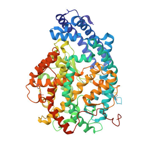
Structural Basis of Ac-Sdkp Hydrolysis by Angiotensin-I Converting Enzyme

Masuyer, G.Douglas, R.G.Sturrock, E.D.Acharya, K.R.

(2015) Sci Rep 5: 13742

  • DOI: https://doi.org/10.1038/srep13742
  • Primary Citation Related Structures: 
    4UFA, 4UFB

  • PubMed Abstract: 

    Angiotensin-I converting enzyme (ACE) is a zinc dipeptidylcarboxypeptidase with two active domains and plays a key role in the regulation of blood pressure and electrolyte homeostasis, making it the principal target in the treatment of cardiovascular disease. More recently, the tetrapetide N-acetyl-Ser-Asp-Lys-Pro (Ac-SDKP) has emerged as a potent antifibrotic agent and negative regulator of haematopoietic stem cell differentiation which is processed exclusively by ACE. Here we provide a detailed biochemical and structural basis for the domain preference of Ac-SDKP. The high resolution crystal structures of N-domain ACE in complex with the dipeptide products of Ac-SDKP cleavage were obtained and offered a template to model the mechanism of substrate recognition of the enzyme. A comprehensive kinetic study of Ac-SDKP and domain co-operation was performed and indicated domain interactions affecting processing of the tetrapeptide substrate. Our results further illustrate the molecular basis for N-domain selectivity and should help design novel ACE inhibitors and Ac-SDKP analogues that could be used in the treatment of fibrosis disorders.


  • Organizational Affiliation
    • Department of Biology and Biochemistry, University of Bath, Claverton Down, Bath BA2 7AY, UK.

Macromolecule Content 

  • Total Structure Weight: 149.8 kDa 
  • Atom Count: 10,977 
  • Modeled Residue Count: 1,214 
  • Deposited Residue Count: 1,258 
  • Unique protein chains: 1

Macromolecules

Find similar proteins by:|  3D Structure
Entity ID: 1
MoleculeChains  Sequence LengthOrganismDetailsImage
ANGIOTENSIN-CONVERTING ENZYME
A, B
629Homo sapiensMutation(s): 8 
EC: 3.2.1 (PDB Primary Data), 3.4.15.1 (PDB Primary Data)
UniProt & NIH Common Fund Data Resources
Find proteins for P12821 (Homo sapiens)
Explore P12821 
Go to UniProtKB:  P12821
PHAROS:  P12821
GTEx:  ENSG00000159640 
Entity Groups
Sequence Clusters30% Identity50% Identity70% Identity90% Identity95% Identity100% Identity
UniProt GroupP12821
Glycosylation
Glycosylation Sites: 3Go to GlyGen: P12821-1
Sequence Annotations
Expand
Reference Sequence

Oligosaccharides

Help  
Entity ID: 2
MoleculeChains Length2D Diagram GlycosylationD Interactions
2-acetamido-2-deoxy-beta-D-glucopyranose-(1-4)-2-acetamido-2-deoxy-beta-D-glucopyranose
C
2N-Glycosylation
Glycosylation Resources
GlyTouCan: G42666HT
GlyCosmos: G42666HT
GlyGen: G42666HT
Entity ID: 3
MoleculeChains Length2D Diagram GlycosylationD Interactions
beta-D-mannopyranose-(1-4)-2-acetamido-2-deoxy-beta-D-glucopyranose-(1-4)-[alpha-L-fucopyranose-(1-6)]2-acetamido-2-deoxy-beta-D-glucopyranose
D
4N-Glycosylation
Glycosylation Resources
GlyTouCan: G32152BH
GlyCosmos: G32152BH
GlyGen: G32152BH
Entity ID: 4
MoleculeChains Length2D Diagram GlycosylationD Interactions
alpha-L-fucopyranose-(1-6)-2-acetamido-2-deoxy-beta-D-glucopyranose
E
2N-Glycosylation
Glycosylation Resources
GlyTouCan: G86851RC
GlyCosmos: G86851RC
GlyGen: G86851RC
Entity ID: 5
MoleculeChains Length2D Diagram GlycosylationD Interactions
beta-D-mannopyranose-(1-4)-2-acetamido-2-deoxy-beta-D-glucopyranose-(1-4)-2-acetamido-2-deoxy-beta-D-glucopyranose
F
3N-Glycosylation
Glycosylation Resources
GlyTouCan: G15407YE
GlyCosmos: G15407YE
GlyGen: G15407YE

Small Molecules

Ligands 7 Unique
IDChains Name / Formula / InChI Key2D Diagram3D Interactions
P6G

Query on P6G


Download:Ideal Coordinates CCD File
L [auth A],
T [auth B],
U [auth B]
HEXAETHYLENE GLYCOL
C12 H26 O7
IIRDTKBZINWQAW-UHFFFAOYSA-N
NAG

Query on NAG


Download:Ideal Coordinates CCD File
I [auth A],
Q [auth B]
2-acetamido-2-deoxy-beta-D-glucopyranose
C8 H15 N O6
OVRNDRQMDRJTHS-FMDGEEDCSA-N
SAC

Query on SAC


Download:Ideal Coordinates CCD File
M [auth A],
V [auth B]
N-ACETYL-SERINE
C5 H9 N O4
JJIHLJJYMXLCOY-BYPYZUCNSA-N
ASP

Query on ASP


Download:Ideal Coordinates CCD File
N [auth A],
W [auth B]
ASPARTIC ACID
C4 H7 N O4
CKLJMWTZIZZHCS-REOHCLBHSA-N
PEG

Query on PEG


Download:Ideal Coordinates CCD File
J [auth A],
K [auth A],
R [auth B],
S [auth B]
DI(HYDROXYETHYL)ETHER
C4 H10 O3
MTHSVFCYNBDYFN-UHFFFAOYSA-N
ZN

Query on ZN


Download:Ideal Coordinates CCD File
G [auth A],
O [auth B]
ZINC ION
Zn
PTFCDOFLOPIGGS-UHFFFAOYSA-N
CL

Query on CL


Download:Ideal Coordinates CCD File
H [auth A],
P [auth B]
CHLORIDE ION
Cl
VEXZGXHMUGYJMC-UHFFFAOYSA-M

Experimental Data & Validation

Experimental Data

  • Method: X-RAY DIFFRACTION
  • Resolution: 1.80 Å
  • R-Value Free:  0.228 (Depositor), 0.228 (DCC) 
  • R-Value Work:  0.202 (Depositor), 0.202 (DCC) 
  • R-Value Observed: 0.204 (Depositor) 
Space Group: P 1
Unit Cell:
Length ( Å )Angle ( ˚ )
a = 72.87α = 88.65
b = 76.72β = 64.31
c = 82.8γ = 75.22
Software Package:
Software NamePurpose
REFMACrefinement
MOSFLMdata reduction
SCALAdata scaling
PHASERphasing

Structure Validation

View Full Validation Report


Ligand Structure Quality Assessment 


Entry History 

Deposition Data

Revision History  (Full details and data files)

  • Version 1.0: 2015-10-07
    Type: Initial release
  • Version 2.0: 2020-07-29
    Type: Remediation
    Reason: Carbohydrate remediation
    Changes: Atomic model, Data collection, Derived calculations, Other, Structure summary
  • Version 2.1: 2023-12-20
    Changes: Data collection, Database references, Refinement description, Structure summary
  • Version 2.2: 2024-11-13
    Changes: Structure summary