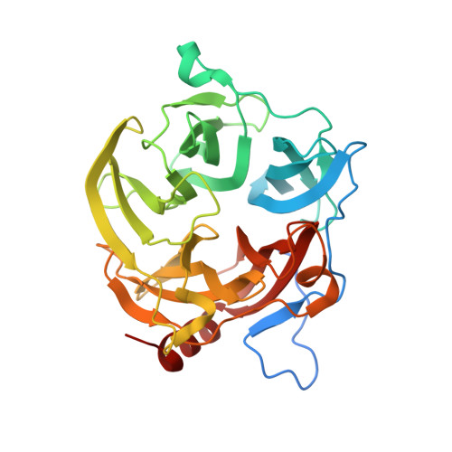
4UDG

Crystal structure of b-1,4-mannopyranosyl-chitobiose phosphorylase at 1.60 Angstrom in complex with N-acetylglucosamine and inorganic phosphate


Experimental Data Snapshot

  • Method: X-RAY DIFFRACTION
  • Resolution: 1.60 Å
  • R-Value Free: 0.183 
  • R-Value Work: 0.154 
  • R-Value Observed: 0.155 

wwPDB Validation   3D Report Full Report


Ligand Structure Quality Assessment 


This is version 2.0 of the entry. See complete history


Literature

Structural Bases for N-Glycan Processing by Mannoside Phosphorylase.

Ladeveze, S.Cioci, G.Roblin, P.Mourey, L.Tranier, S.Potocki-Veronese, G.

(2015) Acta Crystallogr D Biol Crystallogr 71: 1335

  • DOI: https://doi.org/10.1107/S1399004715006604
  • Primary Citation of Related Structures:  
    4UDG, 4UDI, 4UDJ, 4UDK

  • PubMed Abstract: 
  • The first crystal structure of Uhgb_MP, a β-1,4-mannopyranosyl-chitobiose phosphorylase belonging to the GH130 family which is involved in N-glycan degradation by human gut bacteria, was solved at 1.85 Å resolution in the apo form and in complex with mannose and N-acetylglucosamine ...

    The first crystal structure of Uhgb_MP, a β-1,4-mannopyranosyl-chitobiose phosphorylase belonging to the GH130 family which is involved in N-glycan degradation by human gut bacteria, was solved at 1.85 Å resolution in the apo form and in complex with mannose and N-acetylglucosamine. SAXS and crystal structure analysis revealed a hexameric structure, a specific feature of GH130 enzymes among other glycoside phosphorylases. Mapping of the -1 and +1 subsites in the presence of phosphate confirmed the conserved Asp104 as the general acid/base catalytic residue, which is in agreement with a single-step reaction mechanism involving Man O3 assistance for proton transfer. Analysis of this structure, the first to be solved for a member of the GH130_2 subfamily, revealed Met67, Phe203 and the Gly121-Pro125 loop as the main determinants of the specificity of Uhgb_MP and its homologues towards the N-glycan core oligosaccharides and mannan, and the molecular bases of the key role played by GH130 enzymes in the catabolism of dietary fibre and host glycans.


    Organizational Affiliation

    Université de Toulouse; INSA, UPS, INP; LISBP, 135 Avenue de Rangueil, 31077 Toulouse, France.


Macromolecules
Find similar proteins by:  (by identity cutoff)  |  3D Structure
Entity ID: 1
MoleculeChainsSequence LengthOrganismDetailsImage
UHGB_MP
A, B, C, D, E, F
A, B, C, D, E, F
347uncultured organismMutation(s): 0 
EC: 2.4.1
UniProt
Find proteins for D9ZDQ9 (uncultured organism)
Explore D9ZDQ9 
Go to UniProtKB:  D9ZDQ9
Entity Groups  
Sequence Clusters30% Identity50% Identity70% Identity90% Identity95% Identity100% Identity
UniProt GroupD9ZDQ9
Protein Feature View
Expand
  • Reference Sequence
Small Molecules
Ligands 5 Unique
IDChainsName / Formula / InChI Key2D Diagram3D Interactions
NDG
Query on NDG

Download Ideal Coordinates CCD File 
AA [auth E],
GA [auth F],
H [auth A],
N [auth B],
S [auth C],
AA [auth E],
GA [auth F],
H [auth A],
N [auth B],
S [auth C],
W [auth D]
2-acetamido-2-deoxy-alpha-D-glucopyranose
C8 H15 N O6
OVRNDRQMDRJTHS-PVFLNQBWSA-N
 Ligand Interaction
PO4
Query on PO4

Download Ideal Coordinates CCD File 
FA [auth F],
G [auth A],
M [auth B],
R [auth C],
V [auth D],
FA [auth F],
G [auth A],
M [auth B],
R [auth C],
V [auth D],
Z [auth E]
PHOSPHATE ION
O4 P
NBIIXXVUZAFLBC-UHFFFAOYSA-K
 Ligand Interaction
GOL
Query on GOL

Download Ideal Coordinates CCD File 
BA [auth E],
HA [auth F],
I [auth A],
O [auth B],
P [auth B],
BA [auth E],
HA [auth F],
I [auth A],
O [auth B],
P [auth B],
T [auth C],
X [auth D]
GLYCEROL
C3 H8 O3
PEDCQBHIVMGVHV-UHFFFAOYSA-N
 Ligand Interaction
EDO
Query on EDO

Download Ideal Coordinates CCD File 
DA [auth E],
EA [auth E],
JA [auth F],
K [auth A],
L [auth A]
1,2-ETHANEDIOL
C2 H6 O2
LYCAIKOWRPUZTN-UHFFFAOYSA-N
 Ligand Interaction
K
Query on K

Download Ideal Coordinates CCD File 
CA [auth E],
IA [auth F],
J [auth A],
Q [auth B],
U [auth C],
CA [auth E],
IA [auth F],
J [auth A],
Q [auth B],
U [auth C],
Y [auth D]
POTASSIUM ION
K
NPYPAHLBTDXSSS-UHFFFAOYSA-N
 Ligand Interaction
Experimental Data & Validation

Experimental Data

  • Method: X-RAY DIFFRACTION
  • Resolution: 1.60 Å
  • R-Value Free: 0.183 
  • R-Value Work: 0.154 
  • R-Value Observed: 0.155 
  • Space Group: P 21 21 21
Unit Cell:
Length ( Å )Angle ( ˚ )
a = 83.816α = 90
b = 140.912β = 90
c = 168.598γ = 90
Software Package:
Software NamePurpose
REFMACrefinement
XDSdata reduction
XDSdata scaling
PHASERphasing

Structure Validation

View Full Validation Report


Ligand Structure Quality Assessment 


Entry History 

Deposition Data

Revision History  (Full details and data files)

  • Version 1.0: 2015-05-27
    Type: Initial release
  • Version 1.1: 2015-06-17
    Changes: Database references
  • Version 2.0: 2020-07-29
    Type: Remediation
    Reason: Carbohydrate remediation
    Changes: Atomic model, Data collection, Derived calculations, Other, Structure summary