4U97

Crystal Structure of Asymmetric IRAK4 Dimer


Experimental Data Snapshot

  • Method: X-RAY DIFFRACTION
  • Resolution: 2.65 Å
  • R-Value Free: 0.251 
  • R-Value Work: 0.203 
  • R-Value Observed: 0.206 

wwPDB Validation   3D Report Full Report


Ligand Structure Quality Assessment 


This is version 1.1 of the entry. See complete history


Literature

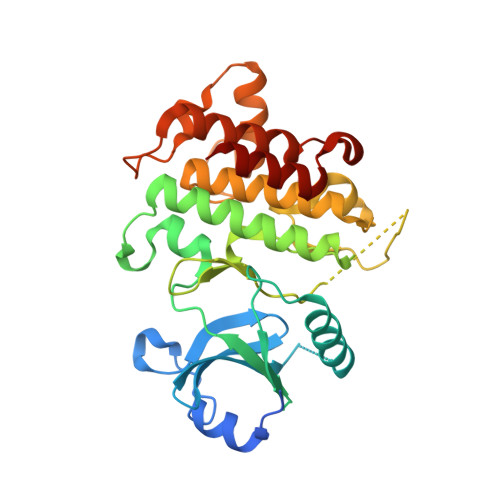
IRAK4 Dimerization and trans-Autophosphorylation Are Induced by Myddosome Assembly.

Ferrao, R.Zhou, H.Shan, Y.Liu, Q.Li, Q.Shaw, D.E.Li, X.Wu, H.

(2014) Mol Cell 55: 891-903

  • DOI: 10.1016/j.molcel.2014.08.006
  • Primary Citation of Related Structures:  
    4U97, 4U9A

  • PubMed Abstract: 
  • Trans-autophosphorylation is among the most prevalent means of protein kinase activation, yet its molecular basis is poorly defined. In Toll-like receptor and interleukin-1 receptor signaling pathways, the kinase IRAK4 is recruited to the membrane-proximal adaptor MyD88 through death domain (DD) interactions, forming the oligomeric Myddosome and mediating NF-κB activation ...

    Trans-autophosphorylation is among the most prevalent means of protein kinase activation, yet its molecular basis is poorly defined. In Toll-like receptor and interleukin-1 receptor signaling pathways, the kinase IRAK4 is recruited to the membrane-proximal adaptor MyD88 through death domain (DD) interactions, forming the oligomeric Myddosome and mediating NF-κB activation. Here we show that unphosphorylated IRAK4 dimerizes in solution with a KD of 2.5 μM and that Myddosome assembly greatly enhances IRAK4 kinase domain (KD) autophosphorylation at sub-KD concentrations. The crystal structure of the unphosphorylated IRAK4(KD) dimer captures a conformation that appears to represent the actual trans-autophosphorylation reaction, with the activation loop phosphosite of one IRAK4 monomer precisely positioned for phosphotransfer by its partner. We show that dimerization is crucial for IRAK4 autophosphorylation in vitro and ligand-dependent signaling in cells. These studies identify a mechanism for oligomerization-driven allosteric autoactivation of IRAK4 that may be general to other kinases activated by autophosphorylation.


    Organizational Affiliation

    Department of Biological Chemistry and Molecular Pharmacology, Harvard Medical School, Boston, MA 02115, USA; Program in Cellular and Molecular Medicine, Boston Children's Hospital, Boston, MA 02115, USA; Weill Cornell Graduate School of Medical Sciences, New York, NY 10065, USA. Electronic address: hao.wu@childrens.harvard.edu.


Macromolecules
Find similar proteins by:  (by identity cutoff)  |  3D Structure
Entity ID: 1
MoleculeChainsSequence LengthOrganismDetailsImage
Interleukin-1 receptor-associated kinase 4A, B312Homo sapiensMutation(s): 1 
Gene Names: IRAK4
EC: 2.7.11.1
UniProt & NIH Common Fund Data Resources
Find proteins for Q9NWZ3 (Homo sapiens)
Explore Q9NWZ3 
Go to UniProtKB:  Q9NWZ3
PHAROS:  Q9NWZ3
Entity Groups  
Sequence Clusters30% Identity50% Identity70% Identity90% Identity95% Identity100% Identity
UniProt GroupQ9NWZ3
Protein Feature View
Expand
  • Reference Sequence
Small Molecules
Ligands 2 Unique
IDChainsName / Formula / InChI Key2D Diagram3D Interactions
STU
Query on STU

Download Ideal Coordinates CCD File 
C [auth A],
D [auth B]
STAUROSPORINE
C28 H26 N4 O3
HKSZLNNOFSGOKW-FYTWVXJKSA-N
 Ligand Interaction
SO4
Query on SO4

Download Ideal Coordinates CCD File 
E [auth B],
F [auth B],
G [auth B]
SULFATE ION
O4 S
QAOWNCQODCNURD-UHFFFAOYSA-L
 Ligand Interaction
Experimental Data & Validation

Experimental Data

  • Method: X-RAY DIFFRACTION
  • Resolution: 2.65 Å
  • R-Value Free: 0.251 
  • R-Value Work: 0.203 
  • R-Value Observed: 0.206 
  • Space Group: P 61 2 2
  • Diffraction Data DOI: 10.15785/SBGRID/98
Unit Cell:
Length ( Å )Angle ( ˚ )
a = 87.58α = 90
b = 87.58β = 90
c = 421.58γ = 120
Software Package:
Software NamePurpose
PHENIXrefinement
XDSdata reduction
XSCALEdata scaling
Cootmodel building

Structure Validation

View Full Validation Report


Ligand Structure Quality Assessment 


Entry History 

Deposition Data

Revision History  (Full details and data files)

  • Version 1.0: 2014-09-24
    Type: Initial release
  • Version 1.1: 2014-10-01
    Changes: Database references